おすすめの製品
由来生物
mouse
品質水準
抗体製品の状態
purified immunoglobulin
抗体製品タイプ
primary antibodies
クローン
DH8.3, monoclonal
交差性
human
包装
antibody small pack of 25 μg
テクニック
ELISA: suitable
flow cytometry: suitable
immunohistochemistry: suitable
immunoprecipitation (IP): suitable
western blot: suitable
アイソタイプ
IgG1κ
NCBIアクセッション番号
UniProtアクセッション番号
ターゲットの翻訳後修飾
unmodified
遺伝子情報
human ... EGFR(1956)
詳細
Epidermal growth factor receptor (UniProt: P00533; also known as EC:2.7.10.1, Proto-oncogene c-ErbB-1, Receptor tyrosine-protein kinase erbB-1) is encoded by the EGFR (also known as ERBB, ERBB1, HER1) gene (Gene ID: 1956) in human. EGFR is a single-pass type 1 membrane protein that is ubiquitously expressed. It is synthesized with a 24 amino acids signal peptide that is subsequently cleaved. It contains an extracellular domain (aa 25-645) a transmembrane domain (aa 646-668), and a cytoplasmic domain (aa 669-1210). Upon binding of EGF, the receptor is phosphorylated and recruits adapter proteins to turn on downstream signaling cascades. EGFR is commonly overexpressed and is mutated in many human malignancies and is often associated with aggressive phenotypes. The most common truncated EGFR is the variant III EGFR deletion mutant (EGFRvIII), containing an inframe deletion of exons 2 7 (801 bp) from the extracellular domain. EGFRvIII mutation is reported in about 5% of human lung squamous cell carcinoma (SCC). It has also been suggested that EGFRvIII potentially plays an essential role in the human NSCLC tumor initiation and maintenance. Drugs such as Gefitinib, erlotnib, and HKI-272 are shown to inhibit the growth of EGFRvIII transformed cells and tumor growth. (Ref.: Ji, H., et al. (2006). Proc. Natl. Acad. Sci. USA 103(20); 7817-7822).
特異性
Clone DH8.3 detects EGFR variant vIII a truncated form of EGFR, present in human cancer cells.
免疫原
KLH-conjugated linear peptide corresponding to EGFRvIII (EGFR variant III) (LEEKKGNYVVTDHC) sequence.
アプリケーション
Research Category
細胞シグナル伝達
細胞シグナル伝達
Anti-EGFRvIII, clone DH8.3, Cat. No. MABS1915, is a mouse monoclonal antibody that specifically detects epidermal growth factor receptor vIIIa and has been tested for use in ELISA, Flow Cytometry, Immunohistochemistry, Immunoprecipitation, and Western Blotting.
Western Blotting Analysis: A representative lot detected EGFRvIII in Western Blotting applications (Lammering, G., et. al. (2004). Clin Cancer Res. 10(19):6732-43; Nishikawa, R., et. al. (2004). Brain Tumor Pathol. 21(2):53-6; Hills, D., et. al. (1995). Int J Cancer. 63(4):537-43).
Flow Cytometry Analysis: A representative lot detected EGFRvIII in Flow Cytometry applications (Johns, T.G., et. al. (2002). Int J Cancer. 98(3):398-408; Hills, D., et. al. (1995). Int J Cancer. 63(4):537-43).
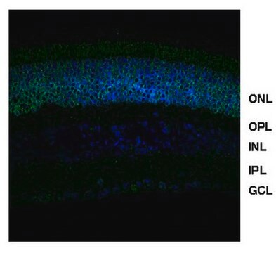
Immunohistochemistry Analysis: A representative lot detected EGFRvIII in Immunohistochemistry applications (Jungbluth, A.A., et. al. (2003). Proc Natl Acad Sci USA. 100(2):639-44; Nishikawa, R., et. al. (2004). Brain Tumor Pathol. 21(2):53-6; Feng, H., et. al. (2014). J Clin Invest. 124(9):3741-56; Feng, H., et. al. (2012). Proc Natl Acad Sci USA. 109(8):3018-23.
Immunoprecipitation Analysis: A representative lot detected EGFRvIII in Immunoprecipitation applications (Lammering, G., et. al. (2004). Clin Cancer Res. 10(19):6732-43; Hills, D., et. al. (1995). Int J Cancer. 63(4):537-43).
ELISA Analysis: A representative lot detected EGFRvIII in ELISA applications (Johns, T.G., et. al. (2002). Int J Cancer. 98(3):398-408; Hills, D., et. al. (1995). Int J Cancer. 63(4):537-43).
品質
Evaluated by Western Blotting in U87MG.∆EGFR cell lysates.
Western Blotting Analysis: 4 ug/mL of this antibody detected EGFRvIII in 10 µg of U87MG.∆EGFR cell lysates.
Western Blotting Analysis: 4 ug/mL of this antibody detected EGFRvIII in 10 µg of U87MG.∆EGFR cell lysates.
ターゲットの説明
~140 kDa observed; 134.28 kDa calculalted. Uncharacterized bands may be observed in some lysate(s).
物理的形状
Protein G purified
Format: Purified
Purified mouse monoclonal antibody IgG1 in buffer containing 0.1 M Tris-Glycine (pH 7.4), 150 mM NaCl with 0.05% sodium azide.
保管および安定性
Stable for 1 year at 2-8°C from date of receipt.
その他情報
Concentration: Please refer to lot specific datasheet.
免責事項
Unless otherwise stated in our catalog or other company documentation accompanying the product(s), our products are intended for research use only and are not to be used for any other purpose, which includes but is not limited to, unauthorized commercial uses, in vitro diagnostic uses, ex vivo or in vivo therapeutic uses or any type of consumption or application to humans or animals.
適切な製品が見つかりませんか。
製品選択ツール.をお試しください
保管分類コード
12 - Non Combustible Liquids
WGK
WGK 1
引火点(°F)
Not applicable
引火点(℃)
Not applicable
適用法令
試験研究用途を考慮した関連法令を主に挙げております。化学物質以外については、一部の情報のみ提供しています。 製品を安全かつ合法的に使用することは、使用者の義務です。最新情報により修正される場合があります。WEBの反映には時間を要することがあるため、適宜SDSをご参照ください。
Jan Code
MABS1915-25UG:
MABS1915:
試験成績書(COA)
製品のロット番号・バッチ番号を入力して、試験成績書(COA) を検索できます。ロット番号・バッチ番号は、製品ラベルに「Lot」または「Batch」に続いて記載されています。
ライフサイエンス、有機合成、材料科学、クロマトグラフィー、分析など、あらゆる分野の研究に経験のあるメンバーがおります。.
製品に関するお問い合わせはこちら(テクニカルサービス)


