おすすめの製品
詳細
Retinoschisin (UniProt: O15537; also known as X-linked juvenile retinoschisis protein) is encoded by the RS1 (also known as XLRS1) gene (Gene ID: 6247) in human. Retinoschisin is discoidin domain-containing protein that is localized along the extracellular surfaces of rod and cone photoreceptors and bipolar cells including the synapses. It is undetectable in the inner plexiform layers and the inner nuclear layer. It is active in cell adhesion process during retinal development and maintains the cellular organization and synaptic structure of the retina. Retinoschisin is a homooctamer of 4 homodimers that are linked by disulfide bonds. It contains a single F5/8 type C domain. Deleterious mutations in RS1 encoding retinoschisin are associated with X-linked juvenile retinoschisis (RS), a common form of macular degeneration in males, which is characterized by a negative electroretinogram pattern and by a splitting of the inner retina. Approximately half of cases of X-linked retinoschisis have bilateral peripheral retinoschisis in the inferotemporal part of the retina. Retinoschisin levels are shown to be up-regulated during the differentiation of a retinoblastoma cell line.
特異性
Clone 3R10 detects Retinoschisin in murine retinal cells. It targets an epitope within 18 amino acids from the N-terminal region.
免疫原
GST-tagged fusion protein containing a the LSSTEDEGEDPWYQKAC peptide fragment corresponding to amino acids 22-39 from the N-terminal region of human Retinoschisin precusor protein.
アプリケーション
Research Category
ニューロサイエンス
ニューロサイエンス
Anti-Retinoschisin, clone 3R10, Cat. No. MABN2434, is a highly specific mouse monoclonal antibody that targets Retinoschisin and has been tested in Immunofluorescence and Western Blotting..
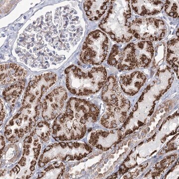
Immunofluorescence Analysis: A representative lot detected Retinoschisin in Immunofluorescence applications (Min, S.H., et. al. (2005). Mol Ther. 12(4):644-51; Weber, B.H., et. al. (2002). Proc Natl Acad Sci USA. 99(9):6222-7).
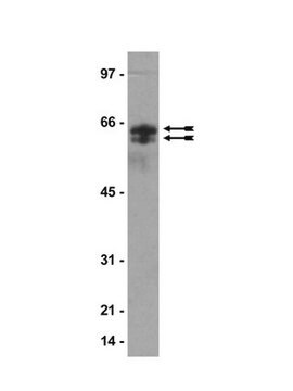
Western Blotting Analysis: A representative lot detected Retinoschisin in Western Blotting applications (Min, S.H., et. al. (2005). Mol Ther. 12(4):644-51; Wu, W.W., et. al. (2003). J Biol Chem. 278(30):28139-46).
Western Blotting Analysis: A representative lot detected Retinoschisin in Western Blotting applications (Min, S.H., et. al. (2005). Mol Ther. 12(4):644-51; Wu, W.W., et. al. (2003). J Biol Chem. 278(30):28139-46).
品質
Evaluated by Western Blotting in mouse retinal tissue lysate.
Western Blotting Analysis: 2 µg/mL of this antibody detected Retinoschisin in 10 µg of mouse retinal tissue lysate.
Western Blotting Analysis: 2 µg/mL of this antibody detected Retinoschisin in 10 µg of mouse retinal tissue lysate.
ターゲットの説明
~23 kDa observed; 25.59 kDa calculated. Uncharacterized bands may be observed in some lysate(s).
物理的形状
Protein G purified
Format: Purified
Purified mouse monoclonal antibody IgG2a in buffer containing 0.1 M Tris-Glycine (pH 7.4), 150 mM NaCl with 0.05% sodium azide.
保管および安定性
Stable for 1 year at 2-8°C from date of receipt.
その他情報
Concentration: Please refer to lot specific datasheet.
免責事項
Unless otherwise stated in our catalog or other company documentation accompanying the product(s), our products are intended for research use only and are not to be used for any other purpose, which includes but is not limited to, unauthorized commercial uses, in vitro diagnostic uses, ex vivo or in vivo therapeutic uses or any type of consumption or application to humans or animals.
適切な製品が見つかりませんか。
製品選択ツール.をお試しください
保管分類コード
12 - Non Combustible Liquids
WGK
WGK 1
引火点(°F)
Not applicable
引火点(℃)
Not applicable
適用法令
試験研究用途を考慮した関連法令を主に挙げております。化学物質以外については、一部の情報のみ提供しています。 製品を安全かつ合法的に使用することは、使用者の義務です。最新情報により修正される場合があります。WEBの反映には時間を要することがあるため、適宜SDSをご参照ください。
Jan Code
MABN2434:
MABN2434-25UG:
試験成績書(COA)
製品のロット番号・バッチ番号を入力して、試験成績書(COA) を検索できます。ロット番号・バッチ番号は、製品ラベルに「Lot」または「Batch」に続いて記載されています。
ライフサイエンス、有機合成、材料科学、クロマトグラフィー、分析など、あらゆる分野の研究に経験のあるメンバーがおります。.
製品に関するお問い合わせはこちら(テクニカルサービス)


