
おすすめの製品


由来生物
mouse
品質水準
抗体製品の状態
purified antibody
抗体製品タイプ
primary antibodies
クローン
HRV-18003, monoclonal
分子量
calculated mol wt 242.824 kDa
observed mol wt ~32 kDa
精製方法
using protein G
交差性
virus
包装
antibody small pack of 100
テクニック
ELISA: suitable
inhibition assay: suitable
western blot: suitable
アイソタイプ
IgG2aκ
エピトープ配列
N-terminal half
タンパク質IDアクセッション番号
UniProtアクセッション番号
保管温度
-10 to -25°C
特異性
Clone HRV-18003 is a mouse monoclonal antibody that detects Rhinovirus. It targets an epitope within 8 amino acids from the N-terminal half.
免疫原
DNA encoding P1-2A and 3C of different strains of Rhinoviruses in pCAGGS plasmids.
アプリケーション
Quality Control Testing
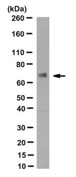
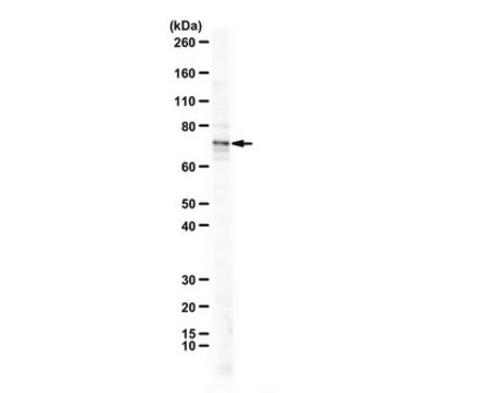
Evaluated by Western Blotting with recombinant Rhinovirus Capsid VP1 protein.
Western Blotting Analysis: A 1:125 dilution of this antibody detected recombinant Rhinovirus Capsid VP1 protein.
Tested Applications
Functional Assay A representative lot of this antibody exhibited strong activity in in vitro antibody dependent cellular phagocytosis (ADCP) assay. (Behzadi, M.A., et al. (2020). Sci Rep.;10(1):9750).
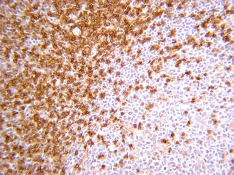
Western Blotting Analysis: A representative lot detected HRV-18003 recognizes VP1 in Western Blotting applications (Behzadi, M.A., et al. (2020). Sci Rep.;10(1):9750).
ELISA Analysis: A representative lot detected HRV-18003 recognizes VP1 in ELISA applications (Behzadi, M.A., et al. (2020). Sci Rep.;10(1):9750).
Note: Actual optimal working dilutions must be determined by end user as specimens, and experimental conditions may vary with the end user.
Evaluated by Western Blotting with recombinant Rhinovirus Capsid VP1 protein.
Western Blotting Analysis: A 1:125 dilution of this antibody detected recombinant Rhinovirus Capsid VP1 protein.
Tested Applications
Functional Assay A representative lot of this antibody exhibited strong activity in in vitro antibody dependent cellular phagocytosis (ADCP) assay. (Behzadi, M.A., et al. (2020). Sci Rep.;10(1):9750).
Western Blotting Analysis: A representative lot detected HRV-18003 recognizes VP1 in Western Blotting applications (Behzadi, M.A., et al. (2020). Sci Rep.;10(1):9750).
ELISA Analysis: A representative lot detected HRV-18003 recognizes VP1 in ELISA applications (Behzadi, M.A., et al. (2020). Sci Rep.;10(1):9750).
Note: Actual optimal working dilutions must be determined by end user as specimens, and experimental conditions may vary with the end user.
ターゲットの説明
Rhinoviruses (RV) belong to the Picornavirus family and are known as a leading cause of respiratory infections. Over 160 different types of rhinoviruses have been identified and based on their genetic diversity and phylogenetic sequence analysis they are grouped as rhinovirus A, B, and C. Three different membrane glycoproteins are known to serve as receptors for these viruses. ICAM-1 is used by a majority of A group and all B group rhinoviruses, a small number of group A viruses use the low-density lipoprotein receptor (LDLR), and the group C of rhinoviruses use cadherin-related family member 3 receptor (CDKR3). Canyons on the surface of the virus are known to provide an attachment site for receptors on the surface of susceptible target cells. Viral infectivity is neutralized when host immunoglobulin G binds to the surface of the virus, thereby blocking interaction between the host cell receptor and the receptor binding site located at the base of the canyon. The RV is composed of single-stranded RNA within a capsid with icosahedral symmetry. The viral capsid of human RV is comprised of four viral proteins: VP1, VP2, VP3, and VP4. The remaining viral proteins are responsible for viral replication and subsequent assembly. Capsid protein VP1 forms an icosahedral capsid of pseudo-T=3 symmetry with capsid proteins VP2 and VP3. VP1 interacts with host cell receptor to provide virion attachment to target host cells. This attachment induces virion internalization. VP1 is the most exposed surface protein and plays a critical role in viral antigenicity and induction of neutralizing antibodies. Clone HRV-18003 binds to a conserve epitope within VP1 and displays cross reactivity with multiple types of rhinoviruses, including A1A, A1B, A15, and A49. It exhibits strong antibody dependent cellular phagocytosis (ADCP) against A1A and A16 viruses. (Ref.: Behzadi, MA., et al. (2020). Sci. Rep. 10(1):9750; Basnet, S., et al. (2019). Chest. ;155(5):1018-1025).
物理的形状
Purified mouse monoclonal antibody IgG2a in PBS without preservatives.
再構成
0.5 mg/mL. Please refer to guidance on suggested starting dilutions and/or titers per application and sample type.
保管および安定性
Store at -10°C to -25°C. Handling Recommendations: Upon receipt and prior to removing the cap, centrifuge the vial and gently mix the solution. Aliquot into microcentrifuge tubes and store at -20°C. Avoid repeated freeze/thaw cycles, which may damage IgG and affect product performance.
その他情報
Concentration: Please refer to the Certificate of Analysis for the lot-specific concentration.
免責事項
Unless otherwise stated in our catalog or other company documentation accompanying the product(s), our products are intended for research use only and are not to be used for any other purpose, which includes but is not limited to, unauthorized commercial uses, in vitro diagnostic uses, ex vivo or in vivo therapeutic uses or any type of consumption or application to humans or animals.
適切な製品が見つかりませんか。
製品選択ツール.をお試しください
保管分類コード
12 - Non Combustible Liquids
WGK
WGK 2
引火点(°F)
Not applicable
引火点(℃)
Not applicable
適用法令
試験研究用途を考慮した関連法令を主に挙げております。化学物質以外については、一部の情報のみ提供しています。 製品を安全かつ合法的に使用することは、使用者の義務です。最新情報により修正される場合があります。WEBの反映には時間を要することがあるため、適宜SDSをご参照ください。
Jan Code
MABF3038-25UG:
MABF3038-100UG:
試験成績書(COA)
製品のロット番号・バッチ番号を入力して、試験成績書(COA) を検索できます。ロット番号・バッチ番号は、製品ラベルに「Lot」または「Batch」に続いて記載されています。
ライフサイエンス、有機合成、材料科学、クロマトグラフィー、分析など、あらゆる分野の研究に経験のあるメンバーがおります。.
製品に関するお問い合わせはこちら(テクニカルサービス)