コンテンツへスキップ
Merck
すべての画像(1)

主要文書

安全性情報

MABF291

Sigma-Aldrich

Anti-S100A8/S100A9 Antibody, clone 5.5

clone 5.5, from mouse

別名:

Protein S100-A9, Calgranulin-B, Calprotectin L1H subunit, Leukocyte L1 complex heavy chain, Migration inhibitory factor-related protein 14, S100 calcium-binding protein A9, AMRP-14, p14, Protein S100-A8, Calgranulin-A, Calprotectin L1L subunit, Cystic fi

ログイン組織・契約価格を表示する


About This Item

UNSPSCコード:
12352203
eCl@ss:
32160702
NACRES:
NA.41

由来生物

mouse

品質水準

抗体製品の状態

purified immunoglobulin

抗体製品タイプ

primary antibodies

クローン

5.5, monoclonal

化学種の反応性

human

テクニック

ELISA: suitable
flow cytometry: suitable
immunohistochemistry: suitable
immunoprecipitation (IP): suitable
western blot: suitable

アイソタイプ

IgG1κ

NCBIアクセッション番号

UniProtアクセッション番号

輸送温度

wet ice

ターゲットの翻訳後修飾

unmodified

遺伝子情報

詳細

S100A8, also known as Protein S100-A8, Calgranulin-A, Calprotectin L1L subunit, Cystic fibrosis antigen (CFAG), Leukocyte L1 complex light chain, Migration inhibitory factor related protein 8 (MRP-8), p8, S100 calcium binding protein A8, or Urinary stone protein band A, and encoded by the gene S100A8/CAGA/CFAG/MRP8, is an important protein that binds both zinc and calcium and plays an important role in the regulation of inflammatory processes and immune response. Found in complex with its partner S100A9, S100A8 facilitates chemotaxis, fatty acid trafficking, cytoskeleton reorganization, and NAPDPH oxidase activation and other intracellular activities, as well as a host of extracellular induced activities such as extracellular proinflammatory, antimicrobial, oxidant scavenging, and apoptotic activities . Additionally S100A8 can act as a potent autoimmunity amplifier and appears to augment various cancers and their spread when over expressed. S100A8 is widely expressed and used as biomarker in patients with inflammatory diseases and as a cancer marker in multiple forms of cancer.

特異性

This antibody detects both S100A8 and S100A9.

免疫原

Whole cells expressing Human S100A8 & S100A9.

アプリケーション

Research Category
炎症及び免疫
Research Sub Category
免疫シグナル伝達
This Anti-S100A8/S100A9 Antibody, clone 5.5 is validated for use in Flow Cytometry and Immunoprecipitation and Immunohistochemistry and ELISA and Western Blotting for the detection of S100A8/S100A9.
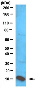
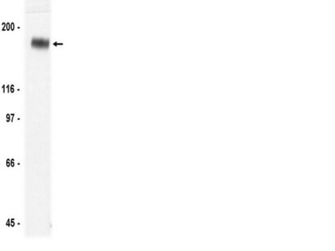
Western Blotting Analysis: A representative lot of this antibody was used to detect S100A8/S100A9 in Human monocyte and neutrophil extracts (Edgeworth, J., et al., (1991) JBC. 266(12):7706-7713).
Western Blotting Analysis: A representative lot of this antibody was used to detect S100A8/S100A9 in neutrophil extracts (Hogg et al., 1989).
Immunoprecipitation Analysis: A representative lot of this antibody was used to detect S100A8/S100A9 in Human monocyte and neutrophil lysate (Edgeworth, J., et al., (1991) JBC. 266(12):7706-7713).
Immunoprecipitation Analysis: A representative lot of this antibody was used to detect S100A8/S100A9 in MRP-8 and TL-14 mutant lysate (Hessian P.A., et al., (2001) Eur. J. Biochem. 268:353-363).
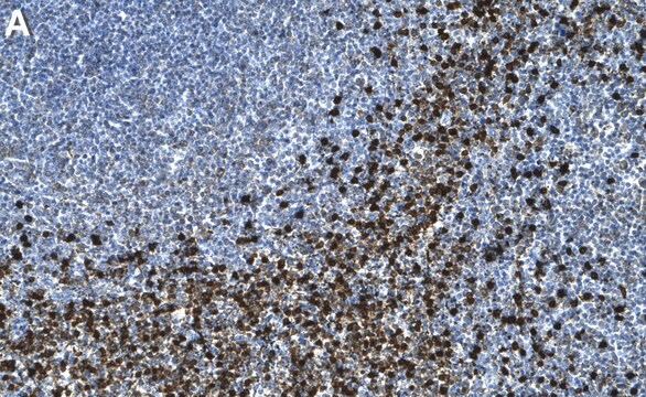
Immunohistochemistry Analysis: A representative lot of this antibody was used to detect S100A8/S100A9 in Human Bronchus tissue (Hogg, N., et al., (1989) Eur. J. Immunol. 19:1053-1061).
Immunohistochemistry Analysis: A representative lot of this antibody was used to detect S100A8/S100A9 in spleen and thymus tissue (Hogg, N., et al., (1989) Eur. J. Immunol. 19:1053-1061).
ELISA: A representative lot of this antibody was used to detect S100A8/S100A9 in ELISA (Ryckman, C., et al., (2003) Arthritis & Rheumatism. 48(8):2310-2320).

品質

Evaluated by Flow Cytometry on Human PBMCs.

Flow Cytometry Analysis: A 1:80 dilution (0.25 µg) of this antibody detected S100A8 and/or S100A9 in 1x10E6 PBMCs.

ターゲットの説明

~8 kda(11kDa) and 14 kDa and as a heterodimer it would be ~24 kDa (under native conditions)

物理的形状

Protein G Purified
Format: Purified
Purified mouse monoclonal IgG1κ in buffer containing 0.1 M Tris-Glycine (pH 7.4), 150 mM NaCl with 0.05% sodium azide.

保管および安定性

Stable for 1 year at 2-8°C from date of receipt.

その他情報

Concentration: Please refer to lot specific datasheet.

免責事項

Unless otherwise stated in our catalog or other company documentation accompanying the product(s), our products are intended for research use only and are not to be used for any other purpose, which includes but is not limited to, unauthorized commercial uses, in vitro diagnostic uses, ex vivo or in vivo therapeutic uses or any type of consumption or application to humans or animals.

適切な製品が見つかりませんか。  

製品選択ツール.をお試しください

保管分類コード

12 - Non Combustible Liquids

WGK

WGK 1

引火点(°F)

Not applicable

引火点(℃)

Not applicable


適用法令

試験研究用途を考慮した関連法令を主に挙げております。化学物質以外については、一部の情報のみ提供しています。 製品を安全かつ合法的に使用することは、使用者の義務です。最新情報により修正される場合があります。WEBの反映には時間を要することがあるため、適宜SDSをご参照ください。

Jan Code

MABF291:


試験成績書(COA)

製品のロット番号・バッチ番号を入力して、試験成績書(COA) を検索できます。ロット番号・バッチ番号は、製品ラベルに「Lot」または「Batch」に続いて記載されています。

以前この製品を購入いただいたことがある場合

文書ライブラリで、最近購入した製品の文書を検索できます。

文書ライブラリにアクセスする

Identification of p8,14 as a highly abundant heterodimeric calcium binding protein complex of myeloid cells.
Edgeworth, J, et al.
The Journal of Biological Chemistry, 266, 7706-7713 (1991)
Monoclonal antibody 5.5 reacts with p8,14, a myeloid molecule associated with some vascular endothelium.
Hogg, N, et al.
European Journal of Immunology, 19, 1053-1061 (1989)
P A Hessian et al.
European journal of biochemistry, 268(2), 353-363 (2001-02-13)
The S100 calcium-binding proteins MRP-8 (S100A8) and MRP-14 (S100A9) form a heterodimeric complex in the cytosol of monocyte and neutrophil cell types circulating in peripheral blood. This complex, but not the individual subunit proteins, is specifically recognized by mAb 27E10.
Carle Ryckman et al.
Arthritis and rheumatism, 48(8), 2310-2320 (2003-08-09)
To examine the role of chemokines, S100A8, and S100A9 in neutrophil accumulation induced by the causative agent of gout, monosodium urate monohydrate (MSU) crystals. MSU crystal-induced neutrophil migration was studied in the murine air-pouch model. Release of chemokines, S100A8, S100A9

ライフサイエンス、有機合成、材料科学、クロマトグラフィー、分析など、あらゆる分野の研究に経験のあるメンバーがおります。.

製品に関するお問い合わせはこちら(テクニカルサービス)