
おすすめの製品
由来生物
mouse
品質水準
抗体製品の状態
purified immunoglobulin
抗体製品タイプ
primary antibodies
クローン
RKSE60, monoclonal
交差性
rat, canine, human, mouse
メーカー/製品名
Chemicon®
テクニック
flow cytometry: suitable
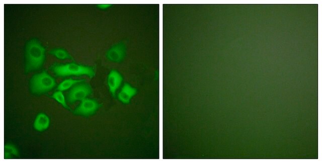
immunocytochemistry: suitable
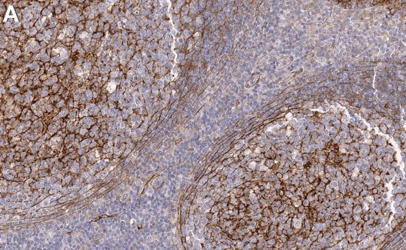
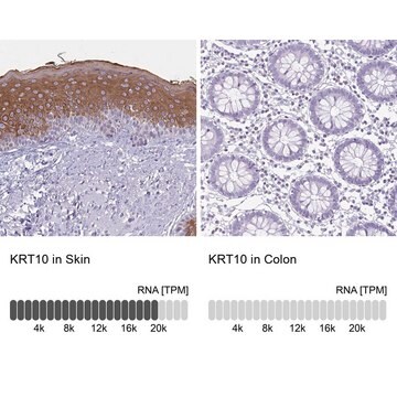
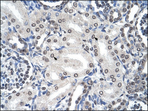
immunohistochemistry: suitable
western blot: suitable
アイソタイプ
IgG1
NCBIアクセッション番号
UniProtアクセッション番号
輸送温度
dry ice
ターゲットの翻訳後修飾
unmodified
遺伝子情報
human ... KRT10(3858)
詳細
Cytokeratins are a subfamily of intermediate filament proteins and are characterized by a remarkable biochemical diversity, represented in epithelial tissues by at least 20 different polypeptides. They range in molecular weight from between 40 kDa and 68 kDa and isoelectric pH between 4.9 - 7.8, The individual cytokeratin polypeptides are designated 1-20. The various epithelia in the human body usually express cytokeratins which are not only characteristic of the type of epithelium, but also related to the degree of maturation or differentiation within an epithelium. Cytokeratin subtype expression patterns are used to an increasing extent in the distinction of different types of epithelial malignancies.
特異性
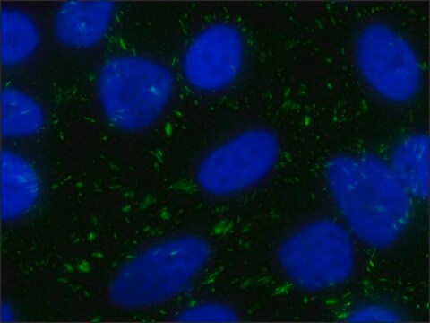
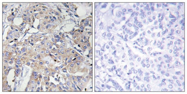
Cytokeratin 10. MAB3230 reacts with keratinizing stratified epithelia and in differentiated areas of highly differentiated squamous cell carcinomas. For the recognition of keratinizing cells in squamous cell carcinomas from epidermis, lung, bladder, cervix, esophagus etc. The antibody reacts only with suprabasal cells of the epidermis and not with basal cells.
免疫原
Cytoskeletal preparation from human squamous keratinizing epithelium.
アプリケーション
Research Category
細胞骨格
細胞骨格
Research Sub Category
サイトケラチン
サイトケラチン
Anti-Cytokeratin 10 Antibody, clone RKSE60 detects level of Cytokeratin 10 & has been published & validated for use in FC, WB, IC, IH.
Western blot
Immunohistochemistry on frozen tissue sections.
Immunocytochemistry
Flow cytometry
Optimal working dilutions must be determined by the end user.
Immunohistochemistry on frozen tissue sections.
Immunocytochemistry
Flow cytometry
Optimal working dilutions must be determined by the end user.
物理的形状
Format: Purified
Liquid in buffer with 0.1% sodium azide.
保管および安定性
Maintain at -20°C in undiluted aliquots up to 6 months. Avoid repeated freeze/thaw cycles.
その他情報
Concentration: Please refer to the Certificate of Analysis for the lot-specific concentration.
法的情報
CHEMICON is a registered trademark of Merck KGaA, Darmstadt, Germany
免責事項
Unless otherwise stated in our catalog or other company documentation accompanying the product(s), our products are intended for research use only and are not to be used for any other purpose, which includes but is not limited to, unauthorized commercial uses, in vitro diagnostic uses, ex vivo or in vivo therapeutic uses or any type of consumption or application to humans or animals.
適切な製品が見つかりませんか。
製品選択ツール.をお試しください
保管分類コード
12 - Non Combustible Liquids
WGK
WGK 2
引火点(°F)
Not applicable
引火点(℃)
Not applicable
適用法令
試験研究用途を考慮した関連法令を主に挙げております。化学物質以外については、一部の情報のみ提供しています。 製品を安全かつ合法的に使用することは、使用者の義務です。最新情報により修正される場合があります。WEBの反映には時間を要することがあるため、適宜SDSをご参照ください。
Jan Code
MAB3230:
試験成績書(COA)
製品のロット番号・バッチ番号を入力して、試験成績書(COA) を検索できます。ロット番号・バッチ番号は、製品ラベルに「Lot」または「Batch」に続いて記載されています。
Loss of nucleoplasmic LAP2alpha-lamin A complexes causes erythroid and epidermal progenitor hyperproliferation.
Nana Naetar,Barbara Korbei,Serguei Kozlov,Marc A Kerenyi,Daniela Dorner,Rosana Kral et al.
Nature Cell Biology null
Atsushi Tokuriki et al.
Carcinogenesis, 30(9), 1645-1650 (2009-07-10)
Inhibitor of DNA binding 2 (Id2) is a negative regulator of basic helix-loop-helix transcription factors and is involved in the control of cellular differentiation and proliferation. By using a two-step chemical carcinogenesis protocol, we evaluated the role of Id2 in
Masashi Miyai et al.
The Journal of investigative dermatology, 136(9), 1848-1857 (2016-05-22)
Mammalian epidermis is a stratified epithelium composed of distinct layers of keratinocytes. The outermost cornified layer is a primary barrier that consists of a cornified envelope, an insoluble structure assembled by cross-linked scaffold proteins, and a surrounding mixture of lipids.
Inflammatory marker analysis in psoriatic skin under topical phosphodiesterase 4 inhibitor treatment.
Lennart M Roesner et al.
The Journal of allergy and clinical immunology, 140(4), 1184-1187 (2017-05-17)
Ryo Ichijo et al.
Nature communications, 8(1), 508-508 (2017-09-13)
The skin surface area varies flexibly in response to body shape changes. Skin homeostasis is maintained by stem cells residing in the basal layer of the interfollicular epidermis. However, how the interfollicular epidermal stem cells response to physiological body shape
ライフサイエンス、有機合成、材料科学、クロマトグラフィー、分析など、あらゆる分野の研究に経験のあるメンバーがおります。.
製品に関するお問い合わせはこちら(テクニカルサービス)