おすすめの製品
由来生物
rabbit
品質水準
抗体製品の状態
affinity isolated antibody
抗体製品タイプ
primary antibodies
クローン
polyclonal
精製方法
affinity chromatography
交差性
rat
交差性(ホモロジーによる予測)
mouse (based on 100% sequence homology), human (based on 100% sequence homology)
テクニック
immunofluorescence: suitable
immunohistochemistry: suitable
western blot: suitable
NCBIアクセッション番号
UniProtアクセッション番号
輸送温度
wet ice
ターゲットの翻訳後修飾
unmodified
遺伝子情報
human ... CELSR1(9620)
詳細
The Flamingo/CELSR family of 7TM-cadherins are characterized with a symmetric protein distribution and polarized activity at neighboring epithelial cell interfaces along defined axes of planar cell polarity. As a component of the extracellular matrix, the CELSR1 protein has been found particularly at the basal surface of neuroepithelial cells within both the early neural tube and a less well-defined group of ventricular zone cells at the midline of the developing spinal cord. CELSR1 plays a role in orienting hair follicles along the anterior–posterior axis of the developing mouse epidermis, both during embryogenesis and postnatal development.
免疫原
Linear peptide corresponding to human CELSR1.
アプリケーション
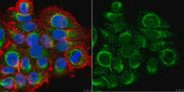
Immunofluorescence Analysis: A 1:500 and 1:1,000 dilution from a representative lot detected CELSR1 in rat cerebellum, substancia nigra, and hippocampus tissue.
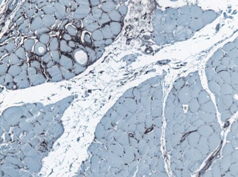
Immunohistochemistry Analysis: A 1:500 dilution from a representative lot detected CELSR1 in neurons of rat cortex tissue and in Purkinje cells and cells within the granular layer of rat cerebellum tissue.
Immunohistochemistry Analysis: A 1:500 dilution from a representative lot detected CELSR1 in neurons of rat cortex tissue and in Purkinje cells and cells within the granular layer of rat cerebellum tissue.
This Anti-CELSR1 Antibody is validated for use in Western Blotting, IHC, Immunofluorescence for the detection of CELSR1.
品質
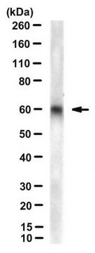
Evaluated by Western Blot in PC12 cell lysate.
Western Blot Analysis: 1 µg/mL of this antibody detected CELSR1 in 10 µg of PC12 cell lysate.
Western Blot Analysis: 1 µg/mL of this antibody detected CELSR1 in 10 µg of PC12 cell lysate.
ターゲットの説明
~330 kDa observed. A second isoform at ~152 kDa may be observed in some lysates.
An uncharacterized band at ~54 kDa may be observed in some cell lysates.
An uncharacterized band at ~54 kDa may be observed in some cell lysates.
その他情報
Concentration: Please refer to the Certificate of Analysis for the lot-specific concentration.
適切な製品が見つかりませんか。
製品選択ツール.をお試しください
保管分類コード
12 - Non Combustible Liquids
WGK
WGK 1
引火点(°F)
Not applicable
引火点(℃)
Not applicable
適用法令
試験研究用途を考慮した関連法令を主に挙げております。化学物質以外については、一部の情報のみ提供しています。 製品を安全かつ合法的に使用することは、使用者の義務です。最新情報により修正される場合があります。WEBの反映には時間を要することがあるため、適宜SDSをご参照ください。
Jan Code
ABT119:
試験成績書(COA)
製品のロット番号・バッチ番号を入力して、試験成績書(COA) を検索できます。ロット番号・バッチ番号は、製品ラベルに「Lot」または「Batch」に続いて記載されています。
Zhengwen An et al.
Nature communications, 9(1), 378-378 (2018-01-27)
The extent to which heterogeneity within mesenchymal stem cell (MSC) populations is related to function is not understood. Using the archetypal MSC in vitro surface marker, CD90/Thy1, here we show that 30% of the MSCs in the continuously growing mouse
ライフサイエンス、有機合成、材料科学、クロマトグラフィー、分析など、あらゆる分野の研究に経験のあるメンバーがおります。.
製品に関するお問い合わせはこちら(テクニカルサービス)