
おすすめの製品


由来生物
mouse
品質水準
抗体製品の状態
purified antibody
抗体製品タイプ
primary antibodies
クローン
LY1C6, monoclonal
フォーム
liquid
含みます
≤0.09% sodium azide as preservative
交差性
rat
メーカー/製品名
Calbiochem®
保管条件
OK to freeze
avoid repeated freeze/thaw cycles
アイソタイプ
IgG1
輸送温度
wet ice
保管温度
−20°C
ターゲットの翻訳後修飾
unmodified
遺伝子情報
rat ... Lamp1(25328)
詳細
免疫原
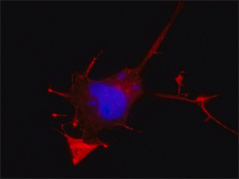
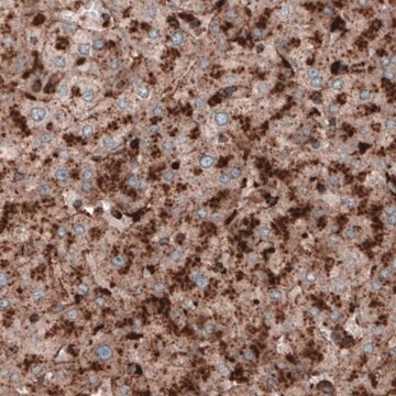
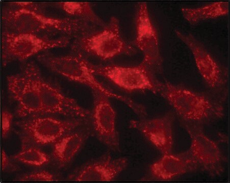
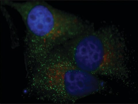
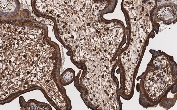
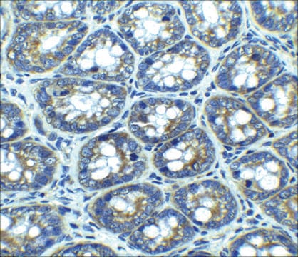
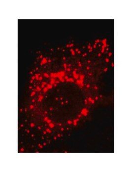
アプリケーション
Immunocytochemistry (1:100)
Immunofluorescence (see comments)
Immunoprecipitation (see comments)
包装
警告
物理的形状
再構成
アナリシスノート
CHO-K1 cells
その他情報
Rohrer, J., et al. 1996. J. Cell Biol. 132, 565.
Howe et al. 1988. PNAS85, 7577.
Lewis et al. 1985. J. Cell Biol.100, 1839.
法的情報
適切な製品が見つかりませんか。
製品選択ツール.をお試しください
保管分類コード
10 - Combustible liquids
WGK
WGK 1
引火点(°F)
Not applicable
引火点(℃)
Not applicable
適用法令
試験研究用途を考慮した関連法令を主に挙げております。化学物質以外については、一部の情報のみ提供しています。 製品を安全かつ合法的に使用することは、使用者の義務です。最新情報により修正される場合があります。WEBの反映には時間を要することがあるため、適宜SDSをご参照ください。
Jan Code
428017-100UG:
428017-UG:
試験成績書(COA)
製品のロット番号・バッチ番号を入力して、試験成績書(COA) を検索できます。ロット番号・バッチ番号は、製品ラベルに「Lot」または「Batch」に続いて記載されています。
資料
Autophagy is a highly regulated process that is involved in cell growth, development, and death. In autophagy cells destroy their own cytoplasmic components in a very systematic manner and recycle them.
Autophagy is a highly regulated process that is involved in cell growth, development, and death. In autophagy cells destroy their own cytoplasmic components in a very systematic manner and recycle them.
ライフサイエンス、有機合成、材料科学、クロマトグラフィー、分析など、あらゆる分野の研究に経験のあるメンバーがおります。.
製品に関するお問い合わせはこちら(テクニカルサービス)