おすすめの製品
由来生物
rabbit
品質水準
クローン
polyclonal
精製方法
affinity chromatography
交差性
mouse, human
メーカー/製品名
ChIPAb+
Upstate®
テクニック
ChIP: suitable
immunocytochemistry: suitable
western blot: suitable
NCBIアクセッション番号
UniProtアクセッション番号
輸送温度
dry ice
遺伝子情報
human ... FOXA1(3169)
詳細
All ChIPAb+ antibodies are individually validated for chromatin precipitation, every lot, every time. Each ChIPAb+ antibody set includes control primers (tested every lot by qPCR) to biologically validate your IP results in a locus-specific context. The qPCR protocol and primer sequences are provided, allowing researchers to validate ChIP protocols when using our antibody in their chromatin context. Each set also includes a negative control antibody to ensure specificity of the ChIP reaction.
The ChIPAb+ FOXA1 set includes the FOXA1 antibody, a Normal Rabbit IgG, and control primers which amplify a 138 bp region of ChIP Primers, Mouse Hnf4α enhancer. The FOXA1 and negative controls are supplied in a scalable "per ChIP" reaction size and can be used to functionally validate the precipitation of FOXA1-associated chromatin.
The ChIPAb+ FOXA1 set includes the FOXA1 antibody, a Normal Rabbit IgG, and control primers which amplify a 138 bp region of ChIP Primers, Mouse Hnf4α enhancer. The FOXA1 and negative controls are supplied in a scalable "per ChIP" reaction size and can be used to functionally validate the precipitation of FOXA1-associated chromatin.
HNF-3A, Hepatocyte nuclear factor 3-alpha (also called FOXA1) is member of the Forkhead family of winged-helix transcription factors. This protein family has around 50 known transcription factors and play an important role in the regulation of cell differentiation, organogenesis, and gene expression. High levels of expression of HNF-3A has been observed in tumors arising from both the prostate and breast. Here it is believed this protein interacts with the androgen receptor in prostate tumors and the estrogen receptor α in breast tumors. In the case of ERα, HNF-3A interacts with cis-regulatory regions in heterochromatin and enhances the interaction of ERα with chromatin.
特異性
This antibody recognizes HNF-3A/FOXA1.
免疫原
Recombinant protein corresponding to the entire protein of human FOXA1
アプリケーション
Research Category
エピジェネティクス及び核内機能分子
エピジェネティクス及び核内機能分子
Research Sub Category
転写因子
転写因子
Chromatin Immunoprecipitation:
Representative lot data.
Sonicated chromatin prepared from Mouse liver tissue (1 mg tissue equivalents per IP) were subjected to chromatin immunoprecipitation using 2 µl of either normal rabbit IgG, or 2 µl Anti-FOXA1 (Part No.CS207368) and the Magna ChIP A Kit (Cat. # 17-610). Successful immunoprecipitation of FoxA1 associated DNA fragments was verified by qPCR using ChIP Primers, Mouse Hnf4α enhancer as a positive locus, and mouse Hnf4α promoter as a negative locus. (Figure 2). Data is presented as percent input of each IP sample relative to input chromatin for each amplicon and ChIP sample as indicated.
Please refer to the EZ-Magna ChIP A (Cat. # 17-408) or EZ-ChIP (Cat. # 17-371) protocol for experimental details.
Western Blotting Analysis:
Representative lot data.
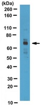
Human pancreas tissue lysate was resolved by electrophoresis, transferred to PVDF membranes and probed with Anti-FOXA1 (0.5 µg/mL).
Proteins were visualized using a Donkey Anti-Rabbit IgG conjugated to HRP and chemiluminescence detection system. (Figure 3)
Arrow indicates FOXA1 (~51 kDa).
Immunocytochemistry Analysis:
Representative lot data.
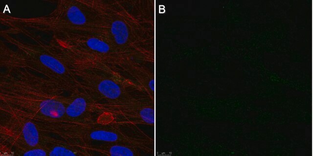
Confocal fluorescent analysis of NIH/3T3 and HeLa cells using Anti-HNF-3A/FOXA1 (Red). Actin filaments have been labeled with Alexa Fluor 488 Dye-Phalloidin (Green). Nucleus is stained with DAPI (Blue). This antibody positively stains nucleus. (Figure 4)
Representative lot data.
Sonicated chromatin prepared from Mouse liver tissue (1 mg tissue equivalents per IP) were subjected to chromatin immunoprecipitation using 2 µl of either normal rabbit IgG, or 2 µl Anti-FOXA1 (Part No.CS207368) and the Magna ChIP A Kit (Cat. # 17-610). Successful immunoprecipitation of FoxA1 associated DNA fragments was verified by qPCR using ChIP Primers, Mouse Hnf4α enhancer as a positive locus, and mouse Hnf4α promoter as a negative locus. (Figure 2). Data is presented as percent input of each IP sample relative to input chromatin for each amplicon and ChIP sample as indicated.
Please refer to the EZ-Magna ChIP A (Cat. # 17-408) or EZ-ChIP (Cat. # 17-371) protocol for experimental details.
Western Blotting Analysis:
Representative lot data.
Human pancreas tissue lysate was resolved by electrophoresis, transferred to PVDF membranes and probed with Anti-FOXA1 (0.5 µg/mL).
Proteins were visualized using a Donkey Anti-Rabbit IgG conjugated to HRP and chemiluminescence detection system. (Figure 3)
Arrow indicates FOXA1 (~51 kDa).
Immunocytochemistry Analysis:
Representative lot data.
Confocal fluorescent analysis of NIH/3T3 and HeLa cells using Anti-HNF-3A/FOXA1 (Red). Actin filaments have been labeled with Alexa Fluor 488 Dye-Phalloidin (Green). Nucleus is stained with DAPI (Blue). This antibody positively stains nucleus. (Figure 4)
This ChIPAb+ FOXA1 -ChIP Validated Antibody & Primer Set conveniently includes the antibody & the specific control PCR primers.
包装
25 assays per set. Recommended use: 2 μL of antibody per chromatin immunoprecipitation (dependent upon biological context).
品質
Chromatin Immunoprecipitation:
Representative lot data
Sonicated chromatin prepared from Mouse liver tissue (1 mg tissue equivalents per IP) were subjected to chromatin immunoprecipitation using 2 µL of either normal rabbit IgG,or 2 µL Anti-FOXA1 and the Magna ChIP® A Kit (Cat. # 17-610). Successful immunoprecipitation of FOXA1- associated DNA fragments was verified by qPCR using ChIP Primers, Mouse Hnf4α enhancer (Figure 1).
Please refer to the EZ-Magna ChIP A (Cat. # 17-408) or EZ-ChIP (Cat. # 17-371) protocol for experimental details.
ターゲットの説明
~51 kDa
物理的形状
Affinity purified
Anti-FOXA1 (Rabbit Polyclonal). One vial containing 50 µL of purified rabbit polyclonal in buffer containing 0.1 M Tris-Glycine (pH 7.4, 150 mM NaCl) with 0.05% sodium azide before the addition of glycerol to 30%. Store at -20°C.
Concentration: 0.35 mg/mL
Normal Rabbit IgG. One vial containing 125 µg Rabbit IgG in 125 µL storage buffer containing 0.05% sodium azide. Store at -20°C.
ChIP Primers, Mouse Hnf4α enhancer. One vial containing 75 μL of 5 μM of each primer specific for Mouse Hnf4α enhancer. Store at -20°C.
FOR: TTC CAG CTG CCT TTA TCT CCC TGT
REV: TCT CCA CAC ATG TCC AGC AGC CT
保管および安定性
Stable for 1 year at -20°C from date of receipt. Handling Recommendations: Upon first thaw, and prior to removing the cap, centrifuge the vial and gently mix the solution. Aliquot into microcentrifuge tubes and store at -20°C. Avoid repeated freeze/thaw cycles, which may damage IgG and affect product performance.
Note: Variability in freezer temperatures below -20°C may cause glycerol containing solutions to become frozen during storage.
Note: Variability in freezer temperatures below -20°C may cause glycerol containing solutions to become frozen during storage.
アナリシスノート
Control
Includes normal rabbit IgG and primers specific for Mouse Hnf4α enhancer.
Includes normal rabbit IgG and primers specific for Mouse Hnf4α enhancer.
その他情報
Concentration: Please refer to the Certificate of Analysis for the lot-specific concentration.
法的情報
MAGNA CHIP is a registered trademark of Merck KGaA, Darmstadt, Germany
UPSTATE is a registered trademark of Merck KGaA, Darmstadt, Germany
免責事項
Unless otherwise stated in our catalog or other company documentation accompanying the product(s), our products are intended for research use only and are not to be used for any other purpose, which includes but is not limited to, unauthorized commercial uses, in vitro diagnostic uses, ex vivo or in vivo therapeutic uses or any type of consumption or application to humans or animals.
保管分類コード
10 - Combustible liquids
適用法令
試験研究用途を考慮した関連法令を主に挙げております。化学物質以外については、一部の情報のみ提供しています。 製品を安全かつ合法的に使用することは、使用者の義務です。最新情報により修正される場合があります。WEBの反映には時間を要することがあるため、適宜SDSをご参照ください。
毒物及び劇物取締法
キットコンポーネントの情報を参照してください
PRTR
キットコンポーネントの情報を参照してください
消防法
キットコンポーネントの情報を参照してください
労働安全衛生法名称等を表示すべき危険物及び有害物
キットコンポーネントの情報を参照してください
労働安全衛生法名称等を通知すべき危険物及び有害物
キットコンポーネントの情報を参照してください
カルタヘナ法
キットコンポーネントの情報を参照してください
Jan Code
キットコンポーネントの情報を参照してください
試験成績書(COA)
製品のロット番号・バッチ番号を入力して、試験成績書(COA) を検索できます。ロット番号・バッチ番号は、製品ラベルに「Lot」または「Batch」に続いて記載されています。
Parisa Mazrooei et al.
Cancer cell, 36(6), 674-689 (2019-11-19)
Thousands of noncoding somatic single-nucleotide variants (SNVs) of unknown function are reported in tumors. Partitioning the genome according to cistromes reveals the enrichment of somatic SNVs in prostate tumors as opposed to adjacent normal tissue cistromes of master transcription regulators
ライフサイエンス、有機合成、材料科学、クロマトグラフィー、分析など、あらゆる分野の研究に経験のあるメンバーがおります。.
製品に関するお問い合わせはこちら(テクニカルサービス)