おすすめの製品
由来生物
rabbit
品質水準
抗体製品の状態
affinity isolated antibody
抗体製品タイプ
primary antibodies
クローン
polyclonal
精製方法
affinity chromatography
化学種の反応性
human
テクニック
immunocytochemistry: suitable
western blot: suitable
NCBIアクセッション番号
UniProtアクセッション番号
輸送温度
wet ice
ターゲットの翻訳後修飾
unmodified
遺伝子情報
human ... CDK5RAP2(55755)
詳細
CDK5RAP2(別名:Cep215)は、多様な細胞内プロセスに関与しています。CDK5R1との相互作用を介して、CDK5活性の潜在的制御因子として働きます。中心小体の解離の負の調節因子として作用し、中心小体の会合と接着を維持します。有糸分裂紡錘体の配向の調節に関与しています。さらに、このタンパク質は、MAD2およびBUBR1プロモーターの転写調節因子として働くことによって、スピンドルチェックポイント活性化を媒介します。CDK5RAP2はMAPRE1と結合して、微小管の重合、束の形成、プラス端での増殖を促進します。また、DNA損傷に誘導されるG2細胞周期チェックポイントにも関与しています。CDK5RAP2は、脳の大きさの減少を特徴とする神経発生疾患である原発性小頭症において変異していることが知られています。
特異性
その他の相同性:ラット(配列相同性83%)およびマウス(配列相同性82%)
免疫原
ヒトCDK5RAP2に対応するMBPタグ付きリコンビナント・タンパク質。
アプリケーション
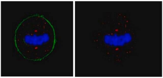
免疫細胞染色:この抗体は、独立した研究所において、RPE1細胞中のCDK5RAP2を検出しました。(Barr, A.R., et al. (2010).J Cell Biol. 189(1):23-39.)。
抗CDK5RAP2抗体(ウサギポリクローナル抗体)は、WBおよびICCにおいて検証済みであり、CDK5RAP2(別名:CDK5調節サブユニット会合タンパク質2、CDK5活性化因子結合タンパク質C48、中心体会合タンパク質215)を検出できます。
品質
HeLaライセート10 µgにおいてウェスタンブロッティングにより評価済み。
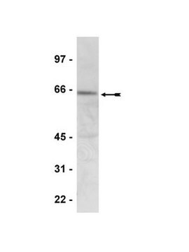
ウェスタンブロッティング:希釈倍率1:2,500で使用、HeLa細胞ライセート10 µg中のCDK5RAP2を検出できます。
ウェスタンブロッティング:希釈倍率1:2,500で使用、HeLa細胞ライセート10 µg中のCDK5RAP2を検出できます。
ターゲットの説明
実測値:約220 kDa。Uniprot.orgには、選択的スプライシングによって産生される4つのアイソフォームが記載されており、これらは一部の細胞ライセートにおいて、分子量約216 kDa、約211 kDa、約210 kDa、約206 kDaで観察される可能性があります。
適切な製品が見つかりませんか。
製品選択ツール.をお試しください
保管分類コード
12 - Non Combustible Liquids
WGK
WGK 1
引火点(°F)
Not applicable
引火点(℃)
Not applicable
適用法令
試験研究用途を考慮した関連法令を主に挙げております。化学物質以外については、一部の情報のみ提供しています。 製品を安全かつ合法的に使用することは、使用者の義務です。最新情報により修正される場合があります。WEBの反映には時間を要することがあるため、適宜SDSをご参照ください。
Jan Code
06-1398:
試験成績書(COA)
製品のロット番号・バッチ番号を入力して、試験成績書(COA) を検索できます。ロット番号・バッチ番号は、製品ラベルに「Lot」または「Batch」に続いて記載されています。
Joo-Hee Sir et al.
The Journal of cell biology, 203(5), 747-756 (2013-12-04)
Most animal cells contain a centrosome, which comprises a pair of centrioles surrounded by an ordered pericentriolar matrix (PCM). Although the role of this organelle in organizing the mitotic spindle poles is well established, its precise contribution to cell division
Andrew Muroyama et al.
The Journal of cell biology, 213(6), 679-692 (2016-06-15)
Differentiation induces the formation of noncentrosomal microtubule arrays in diverse tissues. The formation of these arrays requires loss of microtubule-organizing activity (MTOC) at the centrosome, but the mechanisms regulating this transition remain largely unexplored. Here, we use the robust loss
Alexis R Barr et al.
The Journal of cell biology, 189(1), 23-39 (2010-04-07)
The centrosomal protein, CDK5RAP2, is mutated in primary microcephaly, a neurodevelopmental disorder characterized by reduced brain size. The Drosophila melanogaster homologue of CDK5RAP2, centrosomin (Cnn), maintains the pericentriolar matrix (PCM) around centrioles during mitosis. In this study, we demonstrate a
Tatsuo Miyamoto et al.
Human molecular genetics, 26(22), 4429-4440 (2017-10-04)
Primary microcephaly (MCPH) is an autosomal recessive disorder characterized by congenital reduction of head circumference. Here, we identified compound heterozygous mutations c.731 C > T (p.Ser 244 Leu) and c.2413 G > T (p.Glu 805 X) in the WDR62/MCPH2 gene, which encodes the mitotic centrosomal protein
Shinya Ohta et al.
PloS one, 10(11), e0142798-e0142798 (2015-11-13)
The centrosome-associated C1orf96/Centriole, Cilia and Spindle-Associated Protein (CSAP) targets polyglutamylated tubulin in mitotic microtubules (MTs). Loss of CSAP causes critical defects in brain development; however, it is unclear how CSAP association with MTs affects mitosis progression. In this study, we
ライフサイエンス、有機合成、材料科学、クロマトグラフィー、分析など、あらゆる分野の研究に経験のあるメンバーがおります。.
製品に関するお問い合わせはこちら(テクニカルサービス)


