Passa al contenuto
Merck
Tutte le immagini(4)

Documenti fondamentali

S5768

Sigma-Aldrich

Monoclonal Anti-Synaptophysin antibody produced in mouse

clone SVP-38, ascites fluid

Sinonimo/i:

Synaptophysin Antibody, Synaptophysin Antibody - Monoclonal Anti-Synaptophysin antibody produced in mouse

Autenticatiper visualizzare i prezzi riservati alla tua organizzazione & contrattuali


About This Item

Numero MDL:
Codice UNSPSC:
12352203
NACRES:
NA.41

Origine biologica

mouse

Livello qualitativo

Coniugato

unconjugated

Forma dell’anticorpo

ascites fluid

Tipo di anticorpo

primary antibodies

Clone

SVP-38, monoclonal

PM

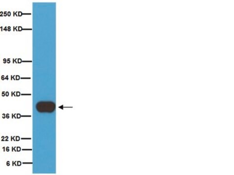
antigen 38 kDa

contiene

15 mM sodium azide

Reattività contro le specie

pig, rat, human, guinea pig

tecniche

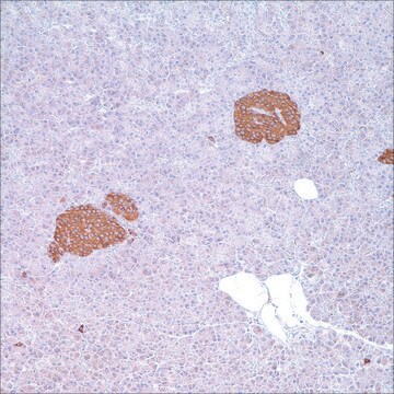
immunohistochemistry (formalin-fixed, paraffin-embedded sections): suitable
immunohistochemistry (frozen sections): 1:200 using rat cerebellum
indirect ELISA: suitable
western blot: suitable

Isotipo

IgG1

N° accesso UniProt

Condizioni di spedizione

dry ice

Temperatura di conservazione

−20°C

modifica post-traduzionali bersaglio

unmodified

Informazioni sul gene

human ... SYP(6855)
rat ... Syp(24804)

Descrizione generale

Synaptophysin (SYP) is an integral membrane glycoprotein of 38,000 Da and it has four membrane-spanning domains. Synaptophysin is localized to neurons, neuromuscular junctions, paraganglia cells, hypophysis, pancreatic islet cells, and adrenal cells. SYP gene is mapped to human chromosome Xp11.23. Monoclonal Anti-Synaptophysin (mouse IgG1 isotype) is derived from the hybridoma produced by the fusion of mouse myeloma cells and splenocytes from an immunized mouse.

Specificità

Labels synaptophysin in neurons, neuromuscular junctions, benign and malignant neural tumors, paraganglia cells, hypophysis, pancreatic islet cells, and adrenal cells.

Immunogeno

Rat retina synaptosome.

Applicazioni

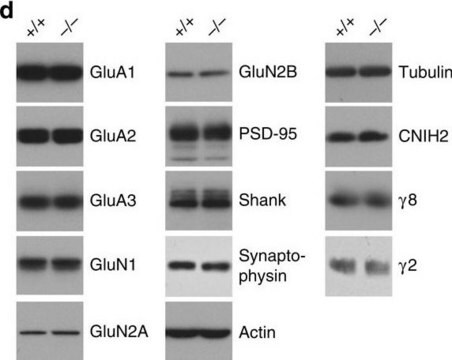
Monoclonal Anti-Synaptophysin antibody has been used in western blotting.

Azioni biochim/fisiol

Synaptophysin participates in calcium dependent exocytosis. The antibody detects synaptophysin in many types of benign and malignant neural and epithelial neuroendocrine neoplasms. It is widely used as a potential marker to study the circulation of synapses in the brain and to discover the basic features of the life cycle of synaptic vesicles (SVs).

Stato fisico

Monoclonal Anti-Synaptophysin is provided as ascites fluid containing 0.1% sodium azide.

Stoccaggio e stabilità

For continuous use, store at 2-8 °C for up to one month. For extended storage, the solution may be frozen in working aliquots. Repeated freezing and thawing is not recommended. Storage in "frost-free" freezers is not recommended. If slight turbidity occurs upon prolonged storage, clarify by centrifugation before use.

Esclusione di responsabilità

Unless otherwise stated in our catalog or other company documentation accompanying the product(s), our products are intended for research use only and are not to be used for any other purpose, which includes but is not limited to, unauthorized commercial uses, in vitro diagnostic uses, ex vivo or in vivo therapeutic uses or any type of consumption or application to humans or animals.

Non trovi il prodotto giusto?  

Prova il nostro Motore di ricerca dei prodotti.

Raccomandato

Codice della classe di stoccaggio

12 - Non Combustible Liquids

Classe di pericolosità dell'acqua (WGK)

WGK 2

Punto d’infiammabilità (°F)

Not applicable

Punto d’infiammabilità (°C)

Not applicable


Scegli una delle versioni più recenti:

Certificati d'analisi (COA)

Lot/Batch Number

Non trovi la versione di tuo interesse?

Se hai bisogno di una versione specifica, puoi cercare il certificato tramite il numero di lotto.

Possiedi già questo prodotto?

I documenti relativi ai prodotti acquistati recentemente sono disponibili nell’Archivio dei documenti.

Visita l’Archivio dei documenti

BDNF-induced synaptic delivery of AMPAR subunits is differentially dependent on NMDA receptors and requires ERK.
Li W and Keifer J
Neurobiology of Learning and Memory, 91(3), 243-249 (2009)
Ken Nakamura et al.
The Journal of neuroscience : the official journal of the Society for Neuroscience, 28(47), 12305-12317 (2008-11-21)
The aggregation of abnormally folded proteins is a defining feature of neurodegenerative disease, but it has not previously been possible to assess the conformation of these proteins in a physiologically relevant context, before they form morphologically recognizable aggregates. We now
Construction of two YAC contigs in human Xp11. 23?p11. 22, one encompassing the loci OATL1, GATA, TFE3, and SYP, the other linking DXS255 to DXS146.
Fisher SE, et al.
Genomics, 29(2), 496-502 (1995)
Coordinate action of pre-and postsynaptic brain-derived neurotrophic factor is required for AMPAR trafficking and acquisition of in vitro classical conditioning.
Li W and Keifer J
Neuroscience, 155(3), 686-697 (2008)
Synaptophysin: leading actor or walk-on role in synaptic vesicle exocytosis?
Valtorta F, et al.
Bioessays, 26(4), 445-453 (2004)

Il team dei nostri ricercatori vanta grande esperienza in tutte le aree della ricerca quali Life Science, scienza dei materiali, sintesi chimica, cromatografia, discipline analitiche, ecc..

Contatta l'Assistenza Tecnica.