Passa al contenuto
Merck
Tutte le immagini(1)

Documenti fondamentali

P2983

Millipore

Anticorpo monoclonale ANTI-FLAG® M2

96-well, clear, polystyrene, flat bottom plate

Sinonimo/i:

Anti-ddddk, Anti-dykddddk

Autenticatiper visualizzare i prezzi riservati alla tua organizzazione & contrattuali


About This Item

Codice UNSPSC:
12352203
NACRES:
NA.32

Tipo di anticorpo

primary antibodies

Livello qualitativo

Materiali

polystyrene

Clone

monoclonal

Durata

Unopened plates are stable for 2 years. Once opened they are stable for 2 weeks.

tecniche

ELISA: suitable

Isotipo

IgG1

Capacità

100-300 ng/well

Temperatura di conservazione

2-8°C

Cerchi prodotti simili? Visita Guida al confronto tra prodotti

Descrizione generale

The Anti-FLAG® HS, M2 Coated Plates are designed to ensure a higher binding capacity than other plates. This is due to the orientation of the Anti-FLAG® M2 mouse monoclonal IgG1 antibody on the plates. The antibody is covalently linked to the surface of a microtiter plate via the Fc portion of the antibody.

Applicazioni

ANTI-FLAG® High Sensitivity, M2 coated 96-well plates is used for Enzyme-linked immunosorbent assays. A convenient, ready to use, platform for the capture and detection of FLAG® fusion proteins. The ANTI-FLAG® M2 antibody is covalently attached to the surface through the Fc portion and can detect 1 ng FLAG fusion protein/well with a capacity of up to 300ng/well. Suitable for screening for expression, study of protein:protein interactions and ELISA assays. Manufactured under ISO 9002 in Sigma′s GMP facility, ANTI-FLAG®P2983 high sensitivity M2 coated multiwell plates utilize a flat bottom, polystyrene baseplate.

Learn more product details in our FLAG® application portal.

Stoccaggio e stabilità

Once plates are opened, they should be stored with desiccant at 2- 8 °C and used within two weeks.

Altre note

Components:
The plate is supplied as a 96-well microtiter plate with clear sides and bottom.
Coating:
ANTI-FLAG® M2 mouse monoclonal antibody, IgG1, is coated at a reaction volume of 200 ml/well.
Blocking:
The wells are pre-blocked for convenience at 275 to 300 ml/well with a complex solution containing bovine serum albumin.
Specificity:
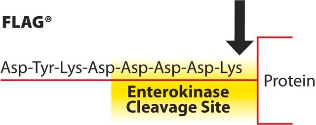
The plates are specific for the FLAG epitope regardless of its placement in the fusion protein: amino-terminal, Met-amino terminal, carboxy terminal or internal. Binding of the epitope is not Ca2+ dependent.
Sensitivity:
Detection of 1 ng/well of a control fusion protein was observed in an ELISA format with p-Nitrophenyl Phosphate (pNPP) as a substrate.
Capacity:
Capture of 100 to 300 ng/well of a FLAG fusion protein has been demonstrated.

Note legali

ANTI-FLAG is a registered trademark of Merck KGaA, Darmstadt, Germany
FLAG is a registered trademark of Merck KGaA, Darmstadt, Germany

Non trovi il prodotto giusto?  

Prova il nostro Motore di ricerca dei prodotti.

Prodotti correlati

N° Catalogo
Descrizione
Determinazione del prezzo

Scegli una delle versioni più recenti:

Certificati d'analisi (COA)

Lot/Batch Number

Non trovi la versione di tuo interesse?

Se hai bisogno di una versione specifica, puoi cercare il certificato tramite il numero di lotto.

Possiedi già questo prodotto?

I documenti relativi ai prodotti acquistati recentemente sono disponibili nell’Archivio dei documenti.

Visita l’Archivio dei documenti

Lilian A Patrón et al.
The Journal of cell biology, 218(3), 993-1010 (2019-01-24)
We genetically characterized the synaptic role of the Drosophila homologue of human DCAF12, a putative cofactor of Cullin4 (Cul4) ubiquitin ligase complexes. Deletion of Drosophila DCAF12 impairs larval locomotion and arrests development. At larval neuromuscular junctions (NMJs), DCAF12 is expressed
He-Wei Jiang et al.
EBioMedicine, 30, 225-236 (2018-04-07)
Owing to the spread of multidrug resistance (MDR) and extensive drug resistance (XDR), there is a pressing need to identify potential targets for the development of more-effective anti-M. tuberculosis (Mtb) drugs. PafA, as the sole Prokaryotic Ubiquitin-like Protein ligase in
Mihaly Mezei et al.
Endocrinology, 161(9) (2020-08-02)
To gain further insight into the binding of the normal and variant human TSHβ subunits (TSHβ and TSHβv), we modeled the 2 monomeric proteins and studied their interaction with the TSH receptor ectodomain (TSHR-ECD) using molecular dynamics simulation Furthermore, analyzed
Natalie F Shanks et al.
The Journal of neuroscience : the official journal of the Society for Neuroscience, 34(36), 12104-12120 (2014-09-05)
Cornichon homologs (CNIHs) are AMPA-type glutamate receptor (AMPAR) auxiliary subunits that modulate AMPAR ion channel function and trafficking. Mechanisms underlying this interaction and functional modulation of the receptor complex are currently unclear. Here, using proteins expressed from mouse and rat
Molecular dissection of the interaction between the AMPA receptor and cornichon homolog-3.
Shanks NF, Cais O, Maruo T, et al.
The Journal of Neuroscience, 34(36), 12104-12120 (2014)

Contenuto correlato

Protein purification techniques, reagents, and protocols for purifying recombinant proteins using methods including, ion-exchange, size-exclusion, and protein affinity chromatography.

Protein expression technologies for expressing recombinant proteins in E. coli, insect, yeast, and mammalian expression systems for fundamental research and the support of therapeutics and vaccine production.

Il team dei nostri ricercatori vanta grande esperienza in tutte le aree della ricerca quali Life Science, scienza dei materiali, sintesi chimica, cromatografia, discipline analitiche, ecc..

Contatta l'Assistenza Tecnica.