Passa al contenuto
Merck
Tutte le immagini(1)

Key Documents

MABE446

Sigma-Aldrich

Anti-Histone H1° Antibody, clone 34

clone 34, from mouse

Sinonimo/i:

Histone H1.0, Histone H1(0), Histone H1°

Autenticatiper visualizzare i prezzi riservati alla tua organizzazione & contrattuali


About This Item

Codice UNSPSC:
12352203
eCl@ss:
32160702
NACRES:
NA.41

Origine biologica

mouse

Livello qualitativo

Forma dell’anticorpo

purified immunoglobulin

Tipo di anticorpo

primary antibodies

Clone

34, monoclonal

Reattività contro le specie

mouse, bovine, Xenopus, human, rat

Reattività contro le specie (prevista in base all’omologia)

ox (immunogen homology)

tecniche

flow cytometry: suitable
immunocytochemistry: suitable
immunohistochemistry: suitable
western blot: suitable

Isotipo

IgG1κ

N° accesso NCBI

N° accesso UniProt

Condizioni di spedizione

wet ice

modifica post-traduzionali bersaglio

unmodified

Informazioni sul gene

human ... H1F0(3005)

Descrizione generale

Histones are highly conserved proteins that serve as the structural scaffold for the organization of nuclear DNA into chromatin. The four core histones, H2A, H2B, H3, and H4, assemble into an octamer (2 molecules of each). Subsequently, 146 base pairs of DNA are wrapped around the octamer, forming a nucleosome. The linker histone, H1, interacts with linker DNA between nucleosomes and functions in the compaction of chromatin into 30nm chromatin fibers and higher order structures.

Specificità

This antibody recognizes Histone H1°.

Immunogeno

Recombinant protein corresponding to Ox liver Histone H1°

Applicazioni

Anti-Histone H1° Antibody, clone 34 is a highly specific mouse monoclonal antibody, that targets Histone H1 & has been tested in western blotting, ICC, IHC & Flow Cytometry.
Research Category
Epigenetics & Nuclear Function
Research Sub Category
Histones
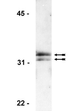
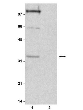
Western Blotting Analysis: A representative lot from an independent laboratory detected Histone H1° in Xenopus embryo tissue lysate (Seigneurin, D., et al. (1995). Int J Dev Biol. 39(4):597-603.; Fu, G., et al. (2003). Biol Reprod. 68(5):1569-1576.).

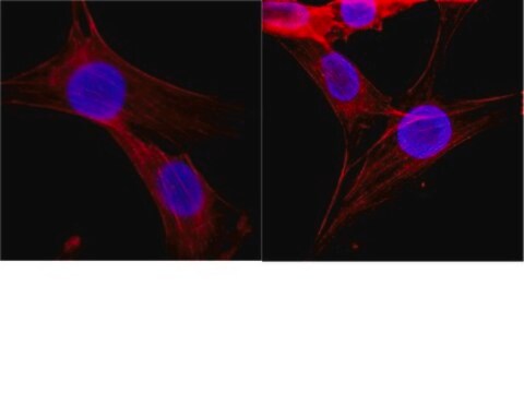
Immunocytochemistry Analyis: A representative lot from an independent laboratory detected Histone H1° in Xenopus unfertilized eggs and early embryos (Fu, G., et al. (2003). Biol Reprod. 68(5):1569-1576.; Adenot, P. G., et al. (2000). J Cell Sci. 113(Pt 16):2897-2907.).

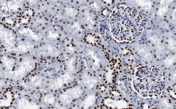
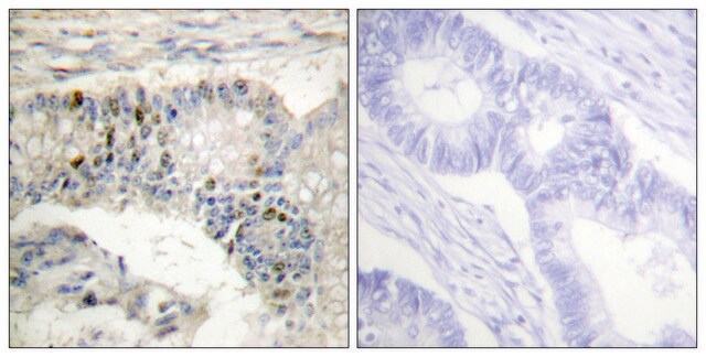
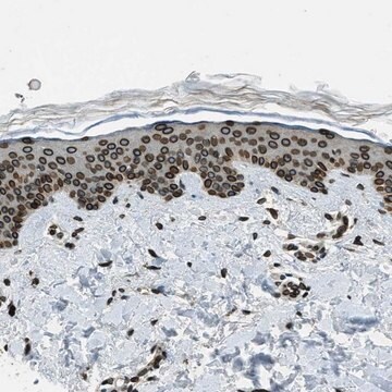
Immunohistochemistry Analysis: A representative lot from an independent laboratory detected Histone H1° in Xenopus embryo tissues (Grunwald, D., et al. (1995). Exp Cell Res. 218(2):586-595.).

Flow Cytometry Analyisis: A representative lot from an independent laboratory detected Histone H1° in FC (Grunwald, D., et al. (1999). Methods Mol Biol. 119:443-454.).

Qualità

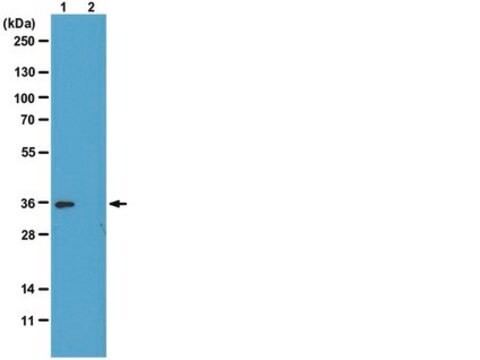
Evaluated by Western Blotting in Jurkat cell lysate.

Western Blotting Analysis: 1 µg/mL of this antibody detected Histone H1° in 10 µg of Jurkat cell lysate.

Descrizione del bersaglio

~30 kDa observed. Uncharacterized band(s) may be observed in some cell lysates.

Stato fisico

Format: Purified
Protein G Purified
Purified mouse monoclonal IgG1κ in buffer containing 0.1 M Tris-Glycine (pH 7.4), 150 mM NaCl with 0.05% sodium azide.

Stoccaggio e stabilità

Stable for 1 year at 2-8°C from date of receipt.

Risultati analitici

Control
Jurkat cell lysate

Altre note

Concentration: Please refer to the Certificate of Analysis for the lot-specific concentration.

Esclusione di responsabilità

Unless otherwise stated in our catalog or other company documentation accompanying the product(s), our products are intended for research use only and are not to be used for any other purpose, which includes but is not limited to, unauthorized commercial uses, in vitro diagnostic uses, ex vivo or in vivo therapeutic uses or any type of consumption or application to humans or animals.

Non trovi il prodotto giusto?  

Prova il nostro Motore di ricerca dei prodotti.

Codice della classe di stoccaggio

12 - Non Combustible Liquids

Classe di pericolosità dell'acqua (WGK)

WGK 1

Punto d’infiammabilità (°F)

Not applicable

Punto d’infiammabilità (°C)

Not applicable


Certificati d'analisi (COA)

Cerca il Certificati d'analisi (COA) digitando il numero di lotto/batch corrispondente. I numeri di lotto o di batch sono stampati sull'etichetta dei prodotti dopo la parola ‘Lotto’ o ‘Batch’.

Possiedi già questo prodotto?

I documenti relativi ai prodotti acquistati recentemente sono disponibili nell’Archivio dei documenti.

Visita l’Archivio dei documenti

Germaine Fu et al.
Biology of reproduction, 68(5), 1569-1576 (2003-02-28)
Oocytes and embryos of many species, including mammals, contain a unique linker (H1) histone, termed H1oo in mammals. It is uncertain, however, whether other H1 histones also contribute to the linker histone complement of these cells. Using immunofluorescence and radiolabeling
D Grunwald et al.
Experimental cell research, 218(2), 586-595 (1995-06-01)
It is known that a transition in the linker-histone variants takes place within chromatin during early development of Xenopus laevis; a cleavage-type H1 is replaced by the somatic type. Based on cytofluorimetric analysis of the distribution of the embryo cells
P G Adenot et al.
Journal of cell science, 113 ( Pt 16), 2897-2907 (2000-07-27)
A striking feature of early embryogenesis in a number of organisms is the use of embryonic linker histones or high mobility group proteins in place of somatic histone H1. The transition in chromatin composition towards somatic H1 appears to be
In situ analysis of chromatin proteins during development and cell differentiation using flow cytometry.
D Grunwald et al.
Methods in molecular biology (Clifton, N.J.), 119, 443-454 (2000-05-11)
Developmentally regulated chromatin acetylation and histone H1(0) accumulation.
Seigneurin, D, et al.
International Journal of Developmental Biology, 39, 597-603 (1995)

Il team dei nostri ricercatori vanta grande esperienza in tutte le aree della ricerca quali Life Science, scienza dei materiali, sintesi chimica, cromatografia, discipline analitiche, ecc..

Contatta l'Assistenza Tecnica.