Pular para o conteúdo
Merck
Todas as fotos(6)

Documentos Principais

HPA040421

Sigma-Aldrich

Anti-NUTM1 antibody produced in rabbit

enhanced validation

Prestige Antibodies® Powered by Atlas Antibodies, affinity isolated antibody, buffered aqueous glycerol solution

Sinônimo(s):

Anti-C15orf55, Anti-Chromosome 15 open reading frame 55, Anti-Dkfzp434o192, Anti-Nut

Faça loginpara ver os preços organizacionais e de contrato


About This Item

Código UNSPSC:
12352203
Número do Atlas de Proteínas Humanas:
NACRES:
NA.41

fonte biológica

rabbit

Nível de qualidade

conjugado

unconjugated

forma do anticorpo

affinity isolated antibody

tipo de produto de anticorpo

primary antibodies

clone

polyclonal

linha de produto

Prestige Antibodies® Powered by Atlas Antibodies

Formulário

buffered aqueous glycerol solution

reatividade de espécies

human

validação aprimorada

orthogonal RNAseq
Learn more about Antibody Enhanced Validation

técnica(s)

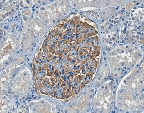
immunohistochemistry: 1:500- 1:1000

sequência de imunogênio

TCPLNVHSYDPQGEGRVDPDLSKPKNLAPLQESQESYTTGTPKATSSHQGLGSTLPRRGTRNAIVPRETSVSKTHRSA

Condições de expedição

wet ice

temperatura de armazenamento

−20°C

modificação pós-traducional do alvo

unmodified

Informações sobre genes

Imunogênio

chromosome 15 open reading frame 55 recombinant protein epitope signature tag (PrEST)

Aplicação

All Prestige Antibodies Powered by Atlas Antibodies are developed and validated by the Human Protein Atlas (HPA) project and as a result, are supported by the most extensive characterization in the industry.

The Human Protein Atlas project can be subdivided into three efforts: Human Tissue Atlas, Cancer Atlas, and Human Cell Atlas. The antibodies that have been generated in support of the Tissue and Cancer Atlas projects have been tested by immunohistochemistry against hundreds of normal and disease tissues and through the recent efforts of the Human Cell Atlas project, many have been characterized by immunofluorescence to map the human proteome not only at the tissue level but now at the subcellular level. These images and the collection of this vast data set can be viewed on the Human Protein Atlas (HPA) site by clicking on the Image Gallery link. We also provide Prestige Antibodies® protocols and other useful information.

Características e benefícios

Prestige Antibodies® are highly characterized and extensively validated antibodies with the added benefit of all available characterization data for each target being accessible via the Human Protein Atlas portal linked just below the product name at the top of this page. The uniqueness and low cross-reactivity of the Prestige Antibodies® to other proteins are due to a thorough selection of antigen regions, affinity purification, and stringent selection. Prestige antigen controls are available for every corresponding Prestige Antibody and can be found in the linkage section.

Every Prestige Antibody is tested in the following ways:
  • IHC tissue array of 44 normal human tissues and 20 of the most common cancer type tissues.
  • Protein array of 364 human recombinant protein fragments.

Ligação

Corresponding Antigen APREST81600

forma física

Solution in phosphate buffered saline, pH 7.2, containing 40% glycerol and 0.02% sodium azide.

Informações legais

Prestige Antibodies is a registered trademark of Merck KGaA, Darmstadt, Germany

Exoneração de responsabilidade

Unless otherwise stated in our catalog or other company documentation accompanying the product(s), our products are intended for research use only and are not to be used for any other purpose, which includes but is not limited to, unauthorized commercial uses, in vitro diagnostic uses, ex vivo or in vivo therapeutic uses or any type of consumption or application to humans or animals.

Não está encontrando o produto certo?  

Experimente o nosso Ferramenta de seleção de produtos.

Código de classe de armazenamento

10 - Combustible liquids

Classe de risco de água (WGK)

WGK 1

Ponto de fulgor (°F)

Not applicable

Ponto de fulgor (°C)

Not applicable


Escolha uma das versões mais recentes:

Certificados de análise (COA)

Lot/Batch Number

Não está vendo a versão correta?

Se precisar de uma versão específica, você pode procurar um certificado específico pelo número do lote ou da remessa.

Já possui este produto?

Encontre a documentação dos produtos que você adquiriu recentemente na biblioteca de documentos.

Visite a Biblioteca de Documentos

Abbas Agaimy et al.
The American journal of surgical pathology, 41(4), 458-471 (2017-03-16)
To more fully characterize the clinical and pathologic spectrum of a recently described tumor entity of the sinonasal tract characterized by loss of nuclear expression of SMARCB1 (INI1), we analyzed 39 SMARCB1-deficient sinonasal carcinomas collected from multiple medical centers. The
Abbas Agaimy et al.
The American journal of surgical pathology, 42(7), 877-884 (2018-04-13)
NUT carcinoma (NC) represents a rare subset of highly aggressive poorly differentiated carcinomas characterized by rearrangement of the NUT (aka NUTM1, nuclear protein in testis) gene, most commonly fused to BRD4. Originally described as a mediastinal/thymic malignancy, NC has been
Cristina Riobello et al.
The American journal of surgical pathology, 44(3), 396-405 (2019-12-27)
A large number of tumor types arise from the mucosa of the sinonasal cavities. Although presenting clinically distinct behavior, due to poorly differentiated histologic features, they can be difficult to classify correctly. Our aim was to investigate whether IDH2 and
Thomas Pincez et al.
Genes, chromosomes & cancer, 59(2), 125-130 (2019-09-14)
Infant acute lymphoblastic leukemias (ALL) are rare hematological malignancies occurring in children younger than 1 year of age, most frequently associated with KMT2A rearrangements (KMT2A-r). The smaller subset without KMT2A-r, which represents 20% of infant ALL cases, is poorly characterized.
Yoon Ah Cho et al.
Thoracic cancer, 11(11), 3205-3212 (2020-10-04)
Nuclear protein in testis (NUT) carcinoma is a rare tumor associated with NUT rearrangement that can present as poorly differentiated to undifferentiated carcinoma, with or without abrupt squamous differentiation. It is often misdiagnosed as poorly differentiated carcinoma or undifferentiated carcinoma

Nossa equipe de cientistas tem experiência em todas as áreas de pesquisa, incluindo Life Sciences, ciência de materiais, síntese química, cromatografia, química analítica e muitas outras.

Entre em contato com a assistência técnica