
44613
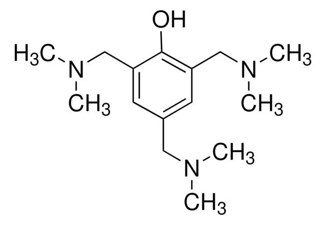
Durcupan™ ACM
single component C, accelerator 960 (DY 060)
Faça loginpara ver os preços organizacionais e de contrato
About This Item
Produtos recomendados


Nível de qualidade
Procurando produtos similares? Visita Guia de comparação de produtos
Aplicação
Embedding material for electron microscopy on the basis of Araldite®.
Informações legais
Araldite is a registered trademark of Huntsman Advanced Materials Inc.
Durcupan is a trademark of Sigma-Aldrich Chemie GmbH
produto relacionado
Nº do produto
Descrição
Preços


Palavra indicadora
Danger
Frases de perigo
Declarações de precaução
Classificações de perigo
Acute Tox. 4 Oral - Eye Dam. 1 - Skin Corr. 1B
Código de classe de armazenamento
8A - Combustible, corrosive hazardous materials
Classe de risco de água (WGK)
WGK 3
Ponto de fulgor (°F)
Not applicable
Ponto de fulgor (°C)
Not applicable
Equipamento de proteção individual
dust mask type N95 (US), Eyeshields, Gloves
Escolha uma das versões mais recentes:
Já possui este produto?
Encontre a documentação dos produtos que você adquiriu recentemente na biblioteca de documentos.
Tin Ki Tsang et al.
eLife, 7 (2018-05-12)
Electron microscopy (EM) offers unparalleled power to study cell substructures at the nanoscale. Cryofixation by high-pressure freezing offers optimal morphological preservation, as it captures cellular structures instantaneously in their near-native state. However, the applicability of cryofixation is limited by its
Daniela Boassa et al.
Cell chemical biology, 26(10), 1407-1416 (2019-08-06)
A protein-fragment complementation assay (PCA) for detecting and localizing intracellular protein-protein interactions (PPIs) was built by bisection of miniSOG, a fluorescent flavoprotein derived from the light, oxygen, voltage (LOV)-2 domain of Arabidopsis phototropin. When brought together by interacting proteins, the
Keun-Young Kim et al.
Cell reports, 29(3), 628-644 (2019-10-17)
The form and synaptic fine structure of melanopsin-expressing retinal ganglion cells, also called intrinsically photosensitive retinal ganglion cells (ipRGCs), were determined using a new membrane-targeted version of a genetic probe for correlated light and electron microscopy (CLEM). ipRGCs project to
David E Gordon et al.
Molecular cell, 78(2), 197-209 (2020-02-23)
We have developed a platform for quantitative genetic interaction mapping using viral infectivity as a functional readout and constructed a viral host-dependency epistasis map (vE-MAP) of 356 human genes linked to HIV function, comprising >63,000 pairwise genetic perturbations. The vE-MAP
Nossa equipe de cientistas tem experiência em todas as áreas de pesquisa, incluindo Life Sciences, ciência de materiais, síntese química, cromatografia, química analítica e muitas outras.
Entre em contato com a assistência técnica