Pular para o conteúdo
Merck
Todas as fotos(1)

Documentos

383A-7

Sigma-Aldrich

SOX-10 Rabbit Polyclonal Antibody

unconjugated, Cell Marque

Faça loginpara ver os preços organizacionais e de contrato


About This Item

Código UNSPSC:
12352203
NACRES:
NA.41

fonte biológica

rabbit

Nível de qualidade

100
500

conjugado

unconjugated

forma do anticorpo

Ig fraction of antiserum

tipo de produto de anticorpo

primary antibodies

clone

polyclonal

descrição

For In Vitro Diagnostic Use in Select Regions (See Chart)

forma

buffered aqueous solution

reatividade de espécies

human

embalagem

vial of 0.1 mL concentrate (383A-74)
vial of 0.5 mL concentrate (383A-75)
bottle of 1.0 mL predilute (383A-77)
vial of 1.0 mL concentrate (383A-76)
bottle of 7.0 mL predilute (383A-78)

fabricante/nome comercial

Cell Marque

técnica(s)

immunohistochemistry (formalin-fixed, paraffin-embedded sections): 1:25-1:100

controle

melanoma, schwannoma, skin melanocytes

Condições de expedição

wet ice

temperatura de armazenamento

2-8°C

visualização

nuclear

Informações sobre genes

human ... SOX10(6663)
mouse ... Sox10(20665)

Descrição geral

Sry-related HMG-BOX gene 10, (SOX-10), a nuclear transcription factor that participates in neural crest development and in the specification and differentiation of cells of melanocytic lineage, has been recently shown to be a sensitive marker of melanoma, including conventional, spindled, and desmoplastic subtypes. SOX-10 nuclear expression was found in 76 of 78 melanomas (97%) and 38 of 77 malignant peripheral nerve sheath tumors (49%), whereas S100 protein was expressed in 71 melanomas (91%) and 23 malignant peripheral nerve sheath tumors (30%). SOX-10 was expressed by metastatic melanomas and nodal capsular nevus in sentinel lymph nodes, but not by other lymph node components such as dendritic cells which usually express S100 protein. It is known that the commonly used melanoma markers, anti-HMB-45 and anti-Melan-A, are poorly expressed in desmoplastic melanomas while it has been reported that anti-SOX-10 was moderately to strongly expressed in all of the 28 desmoplastic melanomas tested. SOX-10 is less likely expressed by background fibrocytes and histiocytes in exicion scar than S100 and MiTF. Therefore, anti-SOX-10 is considered a very reliable marker for recognizing residual demoplastic melanoma.

SOX-10 is diffusely expressed in schwannomas and neurofibromas. SOX-10 reaction is not identified in any other mesenchymal and epithelial tumors except for myoepitheliomas and diffuse astrocytomas. SOX-10 expression is seen in sustentacular cells of pheochromocytomas and paragangliomas, and occasionally carcinoid tumors from various organs, but not in the tumor cells.

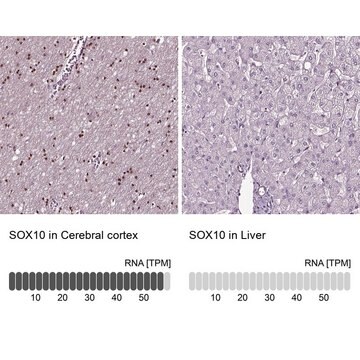
In normal tissues, SOX-10 is expressed in Schwann cells, melanocytes, and myoepithelial cells of salivary, bronchial, eccrine, and mammary glands. SOX-10 expression is also observed in mast cells in a variety of tissues and organs in both nuclear and cytoplasmic reaction.

Qualidade


IVD

IVD

IVD

RUO

Ligação

SOX-10 Positive Control Slides, Product No. 383S, are available for immunohistochemistry (formalin-fixed, paraffin-embedded sections).

forma física

Solution in Tris Buffer, pH 7.3-7.7, with 1% BSA and <0.1% Sodium Azide

Nota de preparo

Download the IFU specific to your product lot and formatNote: This requires a keycode which can be found on your packaging or product label.

Outras notas

For Technical Service please contact: 800-665-7284 or email: service@cellmarque.com

Informações legais

Cell Marque is a trademark of Merck KGaA, Darmstadt, Germany

Not finding the right product?  

Try our Ferramenta de seleção de produtos.

Código de classe de armazenamento

12 - Non Combustible Liquids

Classe de risco de água (WGK)

WGK 2

Ponto de fulgor (°F)

Not applicable

Ponto de fulgor (°C)

Not applicable


Certificados de análise (COA)

Busque Certificados de análise (COA) digitando o Número do Lote do produto. Os números de lote e remessa podem ser encontrados no rótulo de um produto após a palavra “Lot” ou “Batch”.

Já possui este produto?

Encontre a documentação dos produtos que você adquiriu recentemente na biblioteca de documentos.

Visite a Biblioteca de Documentos

A Robson et al.
Histopathology, 38(2), 135-140 (2001-02-24)
The histological distinction of desmoplastic melanoma from cutaneous scar tissue, particularly in the context of re-excision specimens or possible recurrence, may be very difficult. Immunostaining for S100 protein is often used to discriminate although there are little data on S100
Joe A Chorny et al.
The American Journal of dermatopathology, 24(4), 309-312 (2002-07-27)
Distinguishing desmoplastic melanoma (DM) from scar tissue on routine microscopy can be difficult, especially in re-excision specimens, and S100 immunohistochemistry has been recommended as a useful adjunct. The purpose of this study is to evaluate the extent and nature of
Robert N Kelsh
BioEssays : news and reviews in molecular, cellular and developmental biology, 28(8), 788-798 (2006-08-24)
For both vertebrate developmental and evolutionary biologists, and also for clinicians, the neural crest (NC) is a fundamental cell population. An understanding of Sox10 function in NC development is of particular significance since Sox10 mutations underlie several neurocristopathies. Surprisingly, experiments
T A Longacre et al.
The American journal of surgical pathology, 20(12), 1489-1500 (1996-12-01)
The clinical, histologic, and immunohistologic features of 22 desmoplastic melanomas (DMM), 10 mixed desmoplastic and spindle-cell melanomas (DMM/SMM), and two cellular spindle-cell melanomas (SMM) were studied. Patients ranged in age from 35 to 91 years (mean, 67) and included 23
Daisuke Nonaka et al.
The American journal of surgical pathology, 32(9), 1291-1298 (2008-07-19)
S100 protein is a sensitive marker for melanomas and peripheral nerve sheath tumors. It is, however, expressed by other mesenchymal and epithelial tumors. Despite its low specificity, S100 protein is valuable for the diagnosis of desmoplastic melanomas and peripheral nerve

Artigos

Immunohistochemistry (IHC) techniques and applications have greatly improved, dermatopathology is still largely based on H&E stained slides.This paper outlines ways in which IHC antibodies can be utilized for dermatopathology.

Nossa equipe de cientistas tem experiência em todas as áreas de pesquisa, incluindo Life Sciences, ciência de materiais, síntese química, cromatografia, química analítica e muitas outras.

Entre em contato com a assistência técnica