
251R-1
Factor XIIIa (EP3372) Rabbit Monoclonal Primary Antibody
About This Item
Produtos recomendados

fonte biológica
rabbit
Nível de qualidade
100
500
conjugado
unconjugated
forma do anticorpo
culture supernatant
tipo de produto de anticorpo
primary antibodies
clone
EP3372, monoclonal
descrição
For In Vitro Diagnostic Use in Select Regions (See Chart)
Formulário
buffered aqueous solution
reatividade de espécies
human
embalagem
vial of 0.1 mL concentrate (251R-14)
vial of 0.5 mL concentrate (251R-15)
bottle of 1.0 mL predilute (251R-17)
vial of 1.0 mL concentrate (251R-16)
bottle of 7.0 mL predilute (251R-18)
fabricante/nome comercial
Cell Marque®
técnica(s)
immunohistochemistry (formalin-fixed, paraffin-embedded sections): 1:100-1:500
Isotipo
IgG
controle
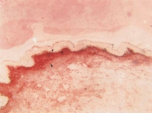
dermatofibroma
Condições de expedição
wet ice
temperatura de armazenamento
2-8°C
visualização
cytoplasmic
Informações sobre genes
human ... F13A1(2162)
Descrição geral
Factor XIIIa is a blood proenzyme that has been identified in platelets, megakaryocyte, and fibroblast-like mesenchymal or histiocytic cells present in the placenta, uterus, and prostate; it is also present in monocytes and macrophages and dermal dendritic cells. Anti- Factor XIIIa has been found to be useful in differentiating between dermatofibroma (90% (+)), dermatofibrosarcoma protuberans (25%(+)) and desmoplastic malignant melanoma (0%(+)). Factor XIIIa positivity is also seen in capillary hemagioblastoma (100%(+)), hemangioendothelioma (100%(+)), hemangiopericytoma (100%(+)), xanthogranuloma (100%(+)), xanthoma (100(+)), hepatocellular carcinoma (93%(+)), glomus tumor (80%(+)), and meningioma (80 % (+)).
Qualidade
 IVD |  IVD |  IVD |  RUO |
Ligação
forma física
Nota de preparo
Outras notas
Informações legais
Não está encontrando o produto certo?
Experimente o nosso Ferramenta de seleção de produtos.
Escolha uma das versões mais recentes:
Certificados de análise (COA)
Não está vendo a versão correta?
Se precisar de uma versão específica, você pode procurar um certificado específico pelo número do lote ou da remessa.
Já possui este produto?
Encontre a documentação dos produtos que você adquiriu recentemente na biblioteca de documentos.
Artigos
Immunohistochemistry (IHC) techniques and applications have greatly improved, dermatopathology is still largely based on H&E stained slides.This paper outlines ways in which IHC antibodies can be utilized for dermatopathology.
Nossa equipe de cientistas tem experiência em todas as áreas de pesquisa, incluindo Life Sciences, ciência de materiais, síntese química, cromatografia, química analítica e muitas outras.
Entre em contato com a assistência técnica