
04511
Live/Dead Cell Double Staining Kit
suitable for fluorescence
Sinônimo(s):
Staining kit for live/dead cells
Faça loginpara ver os preços organizacionais e de contrato
About This Item
Código UNSPSC:
12161503
NACRES:
NA.32
Produtos recomendados


Categorias relacionadas
Aplicação
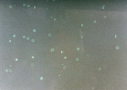
The Live/Dead Cell Double Staining Kit is utilized for simultaneous fluorescence staining of viable and dead cells. This kit contains calcein-AM and propidium iodide (PI) solutions, which stain viable and dead cells, respectively. Calcein-AM, acetoxymethyl ester of calcein, is highly lipophilic and cell membrane permeable. Though calcein-AM itself is not a fluorescent molecule, the calcein generated from Calcein-AM by esterase in a viable cell emits a strong green fluorescence (λex 490 nm, λem 515 nm). Therefore, calcein-AM only stains viable cells. Alternatively, the nuclei staining dye PI cannot pass through a viable cell membrane. It reaches the nucleus by passing through disordered areas of dead cell membrane, and intercalates with the DNA double helix of the cell to emit red fluorescence (λex 535 nm, λem 617 nm). Since both calcein and PI-DNA can be excited with 490 nm light, simultaneous monitoring of viable and dead cells is possible with a fluorescence microscope. Using λex 545 nm, only dead cells can be observed.
Somente componentes do kit
Nº do produto
Descrição
- Solution A (Calcein AM solution) 4 × 50
- Solution B (propidium iodide solution) 300 μL
produto relacionado
Nº do produto
Descrição
Preços
Código de classe de armazenamento
10 - Combustible liquids
Classe de risco de água (WGK)
WGK 2
Ponto de fulgor (°F)
185.0 °F - closed cup
Ponto de fulgor (°C)
85 °C - closed cup
Escolha uma das versões mais recentes:
Já possui este produto?
Encontre a documentação dos produtos que você adquiriu recentemente na biblioteca de documentos.
Os clientes também visualizaram


T Matsuse et al.
Journal of clinical pathology, 51(7), 515-519 (1998-11-03)
To investigate the presence and distribution of advanced glycation end products (AGE) in pulmonary fibrosis. Lung tissue samples obtained from seven necropsy cases with idiopathic pulmonary fibrosis and seven with normal pulmonary parenchyma were examined immunohistochemically with a monoclonal antibody
T Kimura et al.
Neuroscience letters, 208(1), 53-56 (1996-04-12)
The recent immunological demonstration of advanced glycation end products (AGE) of the Maillard reaction in several human tissues suggests a possible involvement of AGE in the aging process. We previously prepared a monoclonal anti-AGE antibody (6D12) which recognized N epsilon-(carboxymethyl)lysine.
T Meshulam et al.
The Journal of infectious diseases, 172(4), 1153-1156 (1995-10-01)
Studies of antimycotic host defenses have been limited by the paucity of rapid, reproducible quantitative assays for fungal cell damage. Prior studies defined a colorimetric method that uses MTT, a tetrazolium dye, to quantify polymorphonuclear leukocyte (PMNL)-mediated damage to fungi.
Elea Boucard et al.
Frontiers in bioengineering and biotechnology, 10, 920929-920929 (2022-08-09)
In tissue engineering, cell origin is important to ensure outcome quality. However, the impact of the cell type chosen for seeding in a biocompatible matrix has been less investigated. Here, we investigated the capacity of primary and immortalized fibroblasts of
S Yoshida et al.
Clinical nephrology, 49(5), 273-280 (1998-06-09)
Cardiovascular disease is one of the most common complications of dialysis and renal transplant patients, and high levels of AGE are present in end-stage renal failure. To address the potential involvement of AGE and growth factors in the pathophysiology of
Artigos
Cell based assays for cell proliferation (BrdU, MTT, WST1), cell viability and cytotoxicity experiments for applications in cancer, neuroscience and stem cell research.
Nossa equipe de cientistas tem experiência em todas as áreas de pesquisa, incluindo Life Sciences, ciência de materiais, síntese química, cromatografia, química analítica e muitas outras.
Entre em contato com a assistência técnica