Pular para o conteúdo
Merck
Todas as fotos(2)

Key Documents

MABN466

Sigma-Aldrich

Anti-Ankyrin-G Antibody, clone N106/36

clone N106/36, from mouse

Sinônimo(s):

Ankyrin-3, ANK-3, Ankyrin-G

Faça loginpara ver os preços organizacionais e de contrato


About This Item

Código UNSPSC:
12352203
eCl@ss:
32160702
NACRES:
NA.41

fonte biológica

mouse

Nível de qualidade

forma do anticorpo

purified immunoglobulin

tipo de produto de anticorpo

primary antibodies

clone

N106/36, monoclonal

reatividade de espécies

rat

técnica(s)

immunohistochemistry: suitable

Isotipo

IgG2aκ

nº de adesão NCBI

nº de adesão UniProt

Condições de expedição

wet ice

modificação pós-traducional do alvo

unmodified

Informações sobre genes

Descrição geral

Ankyrins are a family of spectrin-binding proteins that couple the spectrin/actin cytoskeletal network to the cytoplasmic domains of integral membrane proteins which include channels such as the anion exchanger, voltage-dependent sodium channel, and the Na/K ATPase, as well as neuronal cell adhesion molecules related to L1. In the nervous system, three different ankyrins are known to exist; ankyrinB, ankyrinG, and ankyrinR. All three ankyrins contain an N-terminal membrane binding domain, an internal domain capable of binding spectrin, and a C-terminal regulatory domain which is the target of alternative splicing. AnkyrinB, the predominant neuronal ankyrin includes two isoforms of ~220 kDa and ~440 kDa. The ~220 kDa alternatively spliced isoform is the major form present in the adult brain, while the ~440 kDa form is preferentially expressed in the developing neonatal brain. Three forms of ankyrinG (~190 kDa, ~270 kDa, ~480 kDa) are localized in axonal initial segments and the nodes of Ranvier of myelinated axons where fluxes of action potential occur and ion channels are enriched. Ankyrins comprise ~0.5%-1% of the total membrane protein in the adult vertebrate brain.

Especificidade

Other homologies: Human (92% sequence homology), and Mouse (87% sequence homology).

Imunogênio

Recombinant protein corresponding to human Ankyrin-G.

Aplicação

Anti-Ankyrin-G Antibody, clone N106/36 is a highly specific mouse monoclonal antibody & that targets Ankyrin & has been tested in IHC.
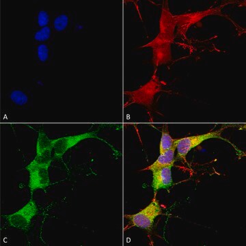
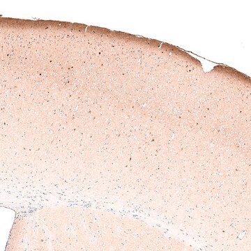
Immunohistochemistry Analysis: A 1:2,000 dilution from a representative lot detected Ankyrin-G in rat cerebral cortex tissue.

Immunohistochemistry Analysis: A representative lot detected Ankyrin-G in rat optic nerve tissue.

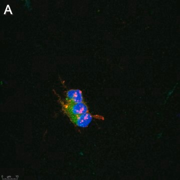
Immunofluorescence Analysis: A representative lot detected Ankyrin-G in adult rat cortex tissue.

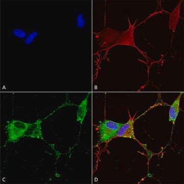
Immunofluorescence Analysis: A representative lot from an independent laboratory detected Ankyrin-G in a rat model of TLE mEC layer II neurons (Hargus, N. J., et al. (2011). Neurobiol Dis. 41(2):361-376.)
Research Category
Neuroscience
Research Sub Category
Developmental Neuroscience

Qualidade

Evaluated by Immunohistochemistry in rat hippocampus tissue.

Immunohistochemistry Analysis: A 1:500 dilution from a representative lot detected Ankyrin-G in rat hippocampus tissue.

Descrição-alvo

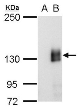
480 kDa calculated

forma física

Format: Purified
Protein G Purified
Purified mouse monoclonal IgG2a κ in 0.1 M Tris-Glycine (pH 7.4), 150 mM NaCl with 0.05% sodium azide.

Armazenamento e estabilidade

Stable for 1 year at 2-8°C from date of receipt.

Nota de análise

Control
Rat hippocampus tissue

Outras notas

Concentration: Please refer to the Certificate of Analysis for the lot-specific concentration.

Exoneração de responsabilidade

Unless otherwise stated in our catalog or other company documentation accompanying the product(s), our products are intended for research use only and are not to be used for any other purpose, which includes but is not limited to, unauthorized commercial uses, in vitro diagnostic uses, ex vivo or in vivo therapeutic uses or any type of consumption or application to humans or animals.

Não está encontrando o produto certo?  

Experimente o nosso Ferramenta de seleção de produtos.

Código de classe de armazenamento

12 - Non Combustible Liquids

Classe de risco de água (WGK)

WGK 1

Ponto de fulgor (°F)

Not applicable

Ponto de fulgor (°C)

Not applicable


Certificados de análise (COA)

Busque Certificados de análise (COA) digitando o Número do Lote do produto. Os números de lote e remessa podem ser encontrados no rótulo de um produto após a palavra “Lot” ou “Batch”.

Já possui este produto?

Encontre a documentação dos produtos que você adquiriu recentemente na biblioteca de documentos.

Visite a Biblioteca de Documentos

Tuancheng Feng et al.
Acta neuropathologica communications, 10(1), 33-33 (2022-03-16)
TMEM106B, a type II lysosomal transmembrane protein, has recently been associated with brain aging, hypomyelinating leukodystrophy, frontotemporal lobar degeneration (FTLD) and several other brain disorders. TMEM106B is critical for proper lysosomal function and TMEM106B deficiency leads to myelination defects, FTLD
Amanda M Brown et al.
Scientific reports, 9(1), 1742-1742 (2019-02-12)
Purkinje cells receive synaptic input from several classes of interneurons. Here, we address the roles of inhibitory molecular layer interneurons in establishing Purkinje cell function in vivo. Using conditional genetics approaches in mice, we compare how the lack of stellate
Kelsie Eichel et al.
Nature, 609(7925), 128-135 (2022-08-18)
Neurons are highly polarized cells that face the fundamental challenge of compartmentalizing a vast and diverse repertoire of proteins in order to function properly1. The axon initial segment (AIS) is a specialized domain that separates a neuron's morphologically, biochemically and
Mable Lam et al.
Nature communications, 13(1), 5583-5583 (2022-09-24)
Myelin is required for rapid nerve signaling and is emerging as a key driver of CNS plasticity and disease. How myelin is built and remodeled remains a fundamental question of neurobiology. Central to myelination is the ability of oligodendrocytes to
Yushan Wan et al.
Cell reports, 33(5), 108346-108346 (2020-11-05)
Complex febrile seizures (FSs) lead to a high risk of intractable temporal lobe epilepsy during adulthood, yet the pathological process of complex FSs is largely unknown. Here, we demonstrate that activated microglia extensively associated with glutamatergic neuronal soma displace surrounding

Nossa equipe de cientistas tem experiência em todas as áreas de pesquisa, incluindo Life Sciences, ciência de materiais, síntese química, cromatografia, química analítica e muitas outras.

Entre em contato com a assistência técnica