
MAB2148-C
Anti-PECAM-1 Antibody, clone P2B1 (Ascites Free)
clone P2B1, 1 mg/mL, from mouse
Sinônimo(s):
Platelet endothelial cell adhesion molecule, PECAM-1, EndoCAM, GPIIA′, PECA1, CD31
About This Item
Produtos recomendados


fonte biológica
mouse
Nível de qualidade
forma do anticorpo
purified antibody
tipo de produto de anticorpo
primary antibodies
clone
P2B1, monoclonal
reatividade de espécies
human
concentração
1 mg/mL
técnica(s)
ELISA: suitable
flow cytometry: suitable
immunocytochemistry: suitable
immunofluorescence: suitable
immunohistochemistry: suitable
immunoprecipitation (IP): suitable
Isotipo
IgG1κ
nº de adesão NCBI
nº de adesão UniProt
Condições de expedição
wet ice
modificação pós-traducional do alvo
unmodified
Informações sobre genes
human ... PECAM1(5175)
Descrição geral
Imunogênio
Aplicação
Immunoprecipitation Analysis: A representative lot detected PECAM-1 by immunoprecipitation.
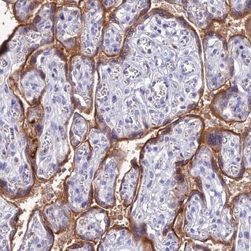
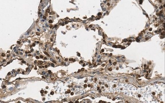
Immunohistochemistry Analysis: A representative lot from an independent laboratory detected PECAM-1 in astrocytoma tissue sections (Bronger, H., et al. (2005). Cancer Res. 65(24):11419-28.).
ELISA Analysis: A representative lot of from an independent laboratory detected PECAM-1 in a panel of CD31 mutant cell lines (Newton, J. P, et al. (1997) J. Biol. Chem., 272: 20555-63.).
Immunofluorescence Analysis: A representative lot from an independent laboratory detected PECAM-1 in astrocytoma tissue sections (Bronger, H., et al. (2005). Cancer Res. 65(24):11419-28.).
Cell Structure
ECM Proteins
Qualidade
Flow Cytometry Analysis: 1 µg of this antibody detected PECAM-1 in 1X10E6 human PBMCs.
Descrição-alvo
Ligação
forma física
Armazenamento e estabilidade
Exoneração de responsabilidade
Não está encontrando o produto certo?
Experimente o nosso Ferramenta de seleção de produtos.
recomendado
Código de classe de armazenamento
12 - Non Combustible Liquids
Classe de risco de água (WGK)
WGK 1
Ponto de fulgor (°F)
Not applicable
Ponto de fulgor (°C)
Not applicable
Certificados de análise (COA)
Busque Certificados de análise (COA) digitando o Número do Lote do produto. Os números de lote e remessa podem ser encontrados no rótulo de um produto após a palavra “Lot” ou “Batch”.
Já possui este produto?
Encontre a documentação dos produtos que você adquiriu recentemente na biblioteca de documentos.
Nossa equipe de cientistas tem experiência em todas as áreas de pesquisa, incluindo Life Sciences, ciência de materiais, síntese química, cromatografia, química analítica e muitas outras.
Entre em contato com a assistência técnica