Pular para o conteúdo
Merck
Todas as fotos(1)

Documentos Principais

MAB2041-I

Sigma-Aldrich

Anti-Laminin β1 Antibody, clone 3E5

Faça loginpara ver os preços organizacionais e de contrato


About This Item

Código UNSPSC:
12352203

fonte biológica

mouse

Nível de qualidade

conjugado

unconjugated

forma do anticorpo

purified antibody

tipo de produto de anticorpo

primary antibodies

clone

3E5, monoclonal

peso molecular

calculated mol wt 198.04 kDa
observed mol wt ~200 kDa

purificado por

using protein G

reatividade de espécies

rat, human

embalagem

antibody small pack of 100 μL

técnica(s)

ELISA: suitable
electron microscopy: suitable
western blot: suitable

Isotipo

IgG

sequência de epítopo

Unknown

nº de adesão de ID de proteína

nº de adesão UniProt

Condições de expedição

dry ice

modificação pós-traducional do alvo

unmodified

Informações sobre genes

Categorias relacionadas

Descrição geral

Laminin subunit b1 (UniProt: P07942; also known as Laminin B1 chain, Laminin-1 subunit beta) is encoded by the LAMB1 gene (Gene ID: 3912) in human. Laminins are the major non-collagenous constituent of basement membranes that participate in wide variety of biological processes, including cell adhesion, differentiation, migration, signaling, neurite outgrowth and metastasis. They are a family of heterotrimeric extracellular glycoproteins consisting of α, β, and γ chains that are bound to each other by disulfide bonds into a cross-shaped molecule comprising one long and three short arms with globules at each end. Laminins mediate the attachment, migration, and organization of cells into tissues during embryonic development by interacting with other extracellular matrix components. Thus far five α, three β, and three γ subunits are described that assemble into at least 15 different laminin molecules. Different α, β, and γ chain isomers combine to give rise to different heterotrimeric laminin isoforms, which are designated by Arabic numerals in the order of their discovery. Laminin- β1 is synthesized with a signal peptide (aa 1-21), which is subsequently cleaved off to generate the mature form. It is expressed in most tissues that produce basement membranes and is one of the 3 chains constituting laminin 1. It is shown to be involved in the organization of the laminar architecture of cerebral cortex and is probably required for the integrity of the basement membrane/glia limitans that serves as an anchor point for the end feet of radial glial cells and as a physical barrier to migrating neurons. (Ref.: Radmanesh, F., et al. (2013). Am. J. Hum. Genet. 92(3); 468-474; Virtanen, I., et al. (1995). J. Histochem. Cytochem. 43(6); 621-628)).
This mouse monoclonal Anti-Laminin β1, clone 3E5, Cat. No. MAB2041-I is tested for use in Western Blotting and ELISA, Inhibition, Electron Microscopy and Western Blotting, for the detection of Laminin subunit β1.

Especificidade

Clone 3E5 is a mouse monoclonal antibody that detects Laminin subunit β1. It targets an epitope near the end of long arm.

Imunogênio

Laminin fragments from human placenta.

Aplicação

Quality Control Testing

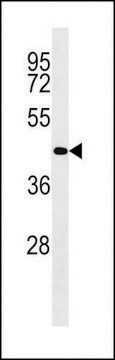
Evaluated by Western Blotting in Human placenta tissue lysates.

Western Blotting Analysis: A 1:500 dilution of this antibody detected Laminin β1 in Human placenta tissue lysates.

Tested Applications

Western Blotting Analysis: A representative lot detected Laminin β1 in Western Blotting applications (Engvall, E., et al. (1986). J Cell Biol.;103(6 Pt1):2457-65).

Electron Microscopy: A representative lot detected Laminin β1 in Electron Microscopy applications (Engvall, E., et al. (1986). J Cell Biol.;103(6 Pt1):2457-65).

Inhibition: A representative lot inhibited the neurite-promoting activity of laminin. (Engvall, E., et al. (1986). J Cell Biol.;103(6 Pt1):2457-65).

ELISA Analysis: A representative lot detected Laminin β1 in ELISA applications (Engvall, E., et al. (1986). J Cell Biol.;103(6 Pt1):2457-65).

Note: Actual optimal working dilutions must be determined by end user as specimens, and experimental conditions may vary with the end user

forma física

Purified mouse monoclonal antibody IgG in PBS without azide.

Armazenamento e estabilidade

Store at -10°C to -25°C. Handling Recommendations: Upon receipt and prior to removing the cap, centrifuge the vial and gently mix the solution. Aliquot into microcentrifuge tubes and store at -20°C. Avoid repeated freeze/thaw cycles, which may damage IgG and affect product performance.

Outras notas

Concentration: Please refer to the Certificate of Analysis for the lot-specific concentration.

Exoneração de responsabilidade

Unless otherwise stated in our catalog or other company documentation accompanying the product(s), our products are intended for research use only and are not to be used for any other purpose, which includes but is not limited to, unauthorized commercial uses, in vitro diagnostic uses, ex vivo or in vivo therapeutic uses or any type of consumption or application to humans or animals.

Não está encontrando o produto certo?  

Experimente o nosso Ferramenta de seleção de produtos.

Código de classe de armazenamento

12 - Non Combustible Liquids

Classe de risco de água (WGK)

WGK 2

Ponto de fulgor (°F)

Not applicable

Ponto de fulgor (°C)

Not applicable


Certificados de análise (COA)

Busque Certificados de análise (COA) digitando o Número do Lote do produto. Os números de lote e remessa podem ser encontrados no rótulo de um produto após a palavra “Lot” ou “Batch”.

Já possui este produto?

Encontre a documentação dos produtos que você adquiriu recentemente na biblioteca de documentos.

Visite a Biblioteca de Documentos

Nossa equipe de cientistas tem experiência em todas as áreas de pesquisa, incluindo Life Sciences, ciência de materiais, síntese química, cromatografia, química analítica e muitas outras.

Entre em contato com a assistência técnica