Pular para o conteúdo
Merck
Todas as fotos(1)

Documentos Principais

MAB1083

Sigma-Aldrich

Anti-GFP Antibody, clone 3F8.2

clone 3F8.2, from mouse

Sinônimo(s):

Green Fluorescent protein

Faça loginpara ver os preços organizacionais e de contrato


About This Item

Código UNSPSC:
12352203
eCl@ss:
32160702
NACRES:
NA.41

fonte biológica

mouse

Nível de qualidade

forma do anticorpo

purified immunoglobulin

tipo de produto de anticorpo

primary antibodies

clone

3F8.2, monoclonal

reatividade de espécies

vertebrates

técnica(s)

western blot: suitable

Isotipo

IgG1κ

nº de adesão UniProt

Condições de expedição

wet ice

modificação pós-traducional do alvo

unmodified

Descrição geral

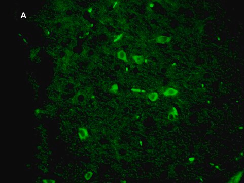
The Green Fluorescent Protein (GFP) from the jellyfish Aequorea victoria is used as a fluorescent indicator for monitoring gene expression in a variety of cellular systems, including living organisms and fixed tissues. Unlike other bioluminescent reporters, GFP fluoresces in the absence of substrates, cofactors, or other intrinsic or extrinsic proteins. Purified GFP is a 27 kDa monomer consisting of 238 amino acids and emits green light (emission maximum at 509 nm) when excited with blue or UV light.

Especificidade

Reacts with most common vertebrate proteins containing the GFP tag.
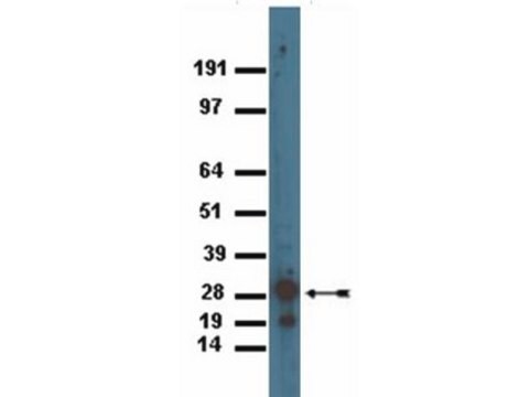
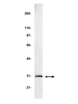
The antibody reacts with the 28 kDa GFP protein.

Imunogênio

Epitope: Full Length
Full length GFP protein.

Aplicação

Detect GFP using this Anti-GFP Antibody, clone 3F8.2 validated for use in WB.
Research Category
Secondary & Control Antibodies
Research Sub Category
Epitope Tags
Western Blot Analysis:
0.125 μg/mL - 1 μg/mL of this antibody detected GFP in 10 ng of GFP recombinant protein (Cat no. 14-392).

Qualidade

Routinely evaluated by Western Blot on GFP recombinant protein.

Descrição-alvo

28 kDa

forma física

Format: Purified
Protein G Purified
Purified mouse immunoglobulin in buffer containing PBS containing 0.05% sodium azide.

Armazenamento e estabilidade

Maintain at 2-8°C in undiluted aliquots for up to 1 year after date of receipt. Avoid repeated freeze/thaw cycles, which may damage IgG and affect product performance.

Nota de análise

Control
Western Blot: GFP recombinant protein

Outras notas

Concentration: Please refer to the Certificate of Analysis for the lot-specific concentration.

Exoneração de responsabilidade

Unless otherwise stated in our catalog or other company documentation accompanying the product(s), our products are intended for research use only and are not to be used for any other purpose, which includes but is not limited to, unauthorized commercial uses, in vitro diagnostic uses, ex vivo or in vivo therapeutic uses or any type of consumption or application to humans or animals.

Não está encontrando o produto certo?  

Experimente o nosso Ferramenta de seleção de produtos.

Código de classe de armazenamento

12 - Non Combustible Liquids

Classe de risco de água (WGK)

WGK 1

Ponto de fulgor (°F)

Not applicable

Ponto de fulgor (°C)

Not applicable


Certificados de análise (COA)

Busque Certificados de análise (COA) digitando o Número do Lote do produto. Os números de lote e remessa podem ser encontrados no rótulo de um produto após a palavra “Lot” ou “Batch”.

Já possui este produto?

Encontre a documentação dos produtos que você adquiriu recentemente na biblioteca de documentos.

Visite a Biblioteca de Documentos

Os clientes também visualizaram

Slide 1 of 2

1 of 2

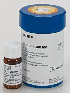
Anti-GFP from mouse IgG1κ (clones 7.1 and 13.1)

Roche

11814460001

Anti-GFP

Protein A Agarose >98% (HPLC and SDS-PAGE), suspension

Roche

PROTAA-RO

Protein A Agarose

Human cardiovascular progenitor cells develop from a KDR+ embryonic-stem-cell-derived population.
Yang, Lei, et al.
Nature, 453, 524-528 (2008)
Bernadette M DeRussy et al.
Journal of virology, 90(16), 7109-7117 (2016-05-27)
Human cytomegalovirus (HCMV) pUL93 and pUL77 are both essential for virus growth, but their functions in the virus life cycle remain mostly unresolved. Homologs of pUL93 and pUL77 in herpes simplex virus 1 (HSV-1) and pseudorabies virus (PRV) are known
Jake D Howden et al.
BMC biology, 19(1), 130-130 (2021-06-24)
Keratinocytes form the main protective barrier in the skin to separate the underlying tissue from the external environment. In order to maintain this barrier, keratinocytes form robust junctions between neighbouring cells as well as with the underlying extracellular matrix. Cell-cell
Markus S Spurlock et al.
Journal of neurotrauma, 34(11), 1981-1995 (2017-03-03)
Penetrating traumatic brain injury (PTBI) is one of the major cause of death and disability worldwide. Previous studies with penetrating ballistic-like brain injury (PBBI), a PTBI rat model revealed widespread perilesional neurodegeneration, similar to that seen in humans following gunshot

Nossa equipe de cientistas tem experiência em todas as áreas de pesquisa, incluindo Life Sciences, ciência de materiais, síntese química, cromatografia, química analítica e muitas outras.

Entre em contato com a assistência técnica