Skip to Content
MilliporeSigma
All Photos(1)

Key Documents

T3413

Sigma-Aldrich

Monoclonal Anti-Tenascin antibody produced in rat

clone MTn-12, ascites fluid

Synonym(s):

Anti-Tenascin-N

Sign Into View Organizational & Contract Pricing


About This Item

MDL number:
UNSPSC Code:
12352203
NACRES:
NA.41

biological source

rat

Quality Level

conjugate

unconjugated

antibody form

ascites fluid

antibody product type

primary antibodies

clone

MTn-12, monoclonal

contains

15 mM sodium azide

species reactivity

mouse

technique(s)

immunohistochemistry (frozen sections): suitable
immunoprecipitation (IP): suitable
indirect ELISA: suitable
indirect immunofluorescence: 1:200 using unfixed, frozen tissue sections of mouse intestine
western blot: suitable

isotype

IgG1

UniProt accession no.

shipped in

dry ice

storage temp.

−20°C

target post-translational modification

unmodified

Gene Information

mouse ... Tnn(329278)

General description

Monoclonal Anti-Mouse Tenascin (rat IgG1 isotype) is derived from the MTn-12 hybridoma1 produced by the fusion of rat myeloma cells and splenocytes from a Lou rat immunized with partially purified mouse tenascin. Human tenascin has three subunits of 190, 200 and 220 kDa. Tenascin has been independently discovered in a variety of species and tissue types, often in the basement membrane or intercellular spaces. It has been described under a variety of names: cytotactin, hexabrachion protein, J1, myotendinous antigen (MI) and glioma mesenchymal extracellular matrix (GMEM). The tenascin molecule is a disulfide-linked hexamer, depending on species, the molecular weights of the subunits range from 190 to 320 kDa.
Tenascin is a high molecular weight, multifunctional, extracellular matrix glycoprotein expressed in association with mesenchymal-epithelial interactions during development and in the neovasculature and stroma of undifferentiated tumors. It has been described under a variety of names: cytotactin, hexabrachion protein, J1, myotendinous antigen (MI) and glioma mesenchymal extracellular matrix (GMEM).
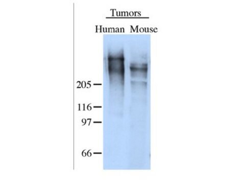
The tenascin molecule is a disulfide-linked hexamer; depending on species, the molecular weights of the subunits range from 190 to 320 kDa. In the mouse, two major subunits of tenascin with an apparent molecular weight of 210 and 260 kDa have been described. The shorter polypeptide predominates during earlier developmental stages and the larger polypeptide appears later in the embryonic gut and especially in the adult intestine. The expression of tenascin is associated with development and growth, both normal and pathological, whereas the distribution in normal adult tissue is restricted. It was proposed that actively growing, migrating and differentiating epithelial sheets can produce factors that can stimulate tenascin expression in the nearby mesenchyme. Human and chicken tenascin contain an RGD sequence which may function in cell adhesion and it seems likely that tenascin mediates cell attachment through an RGD dependent integrin receptor.

Specificity

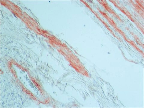
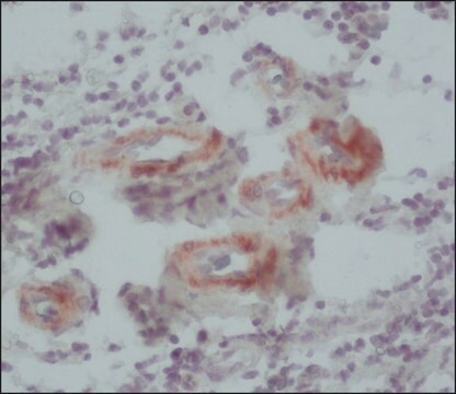
The antibody localizes mouse tenascin in the supernatant of cultured mouse fibroblasts and tissue extracts. No cross-reactivity with tenascin of other species has been observed. In immunohistological testing of frozen tissue sections of mouse intestine, the antibody labels the core of the villi, but not the epithelial cells.

Immunogen

partially purified mouse tenascin.

Application

Applications in which this antibody has been used successfully, and the associated peer-reviewed papers, are given below.
Immunohistochemistry (1 paper)
Monoclonal Anti-Mouse Tenascin antibody may be used for the localization of tenascin and to study of the role of tenascin in epithelial-mesenchymal interactions using various immunochemical assays including ELISA, immunoblot, dot blot and immunohistology.
Monoclonal Anti-Tenascin antibody produced in rat has been used in:
  • Enzyme linked immunosorbent assay (ELISA)
  • Dot blot.
  • Immunoblotting
  • Fluorescence microscopy and immunostaining
  • Immunofluorescence
  • Immunohistochemistry

Biochem/physiol Actions

Tenascin is a high molecular weight, multifunctional, extracellular matrix glycoprotein expressed in association with mesenchymal-epithelial interactions during development and in the neovasculature and stroma of undifferentiated tumors. The expression of tenascin is associated with development and growth, both normal and pathological, whereas the distribution in normal adult tissue is restricted. It was proposed that actively growing, migrating and differentiating epithelial sheets can produce factors that can stimulate tenascin expression in the nearby mesenchyme. Human and chicken tenascin contain an RGD sequence motif which may function in cell adhesion and may be recognized by integrin receptor.

Disclaimer

Unless otherwise stated in our catalog or other company documentation accompanying the product(s), our products are intended for research use only and are not to be used for any other purpose, which includes but is not limited to, unauthorized commercial uses, in vitro diagnostic uses, ex vivo or in vivo therapeutic uses or any type of consumption or application to humans or animals.

Not finding the right product?  

Try our Product Selector Tool.

Storage Class

10 - Combustible liquids

wgk_germany

nwg

flash_point_f

Not applicable

flash_point_c

Not applicable


Certificates of Analysis (COA)

Search for Certificates of Analysis (COA) by entering the products Lot/Batch Number. Lot and Batch Numbers can be found on a product’s label following the words ‘Lot’ or ‘Batch’.

Already Own This Product?

Find documentation for the products that you have recently purchased in the Document Library.

Visit the Document Library

Developmental changes in patterns of expression of tenascin-C variants in the human cornea
Maseruka H, et al.
Investigative Ophthalmology & Visual Science, 41(13), 4101-4107 (2000)
Tumor-derived Tenascin-C Promotes the Epithelial-Mesenchymal Transition in Colorectal Cancer Cells
Takahashi Y, et al.
Anticancer Research, 33(5), 1927-1934 (2013)
Clinical impact and functional aspects of tenascin-C expression during glioma progression
Herold-Mende C, et al.
International Journal of Cancer. Journal International Du Cancer, 98(3), 362-369 (2002)
Site-specific HNK-1 epitope on alternatively spliced fibronectin type-III repeats in tenascin-C promotes neurite outgrowth of hippocampal neurons through contactin-1
Nakamura A, et al.
Testing, 14(1), e0210193-e0210193 (2019)
Tumor targeting by an aptamer
Hicke BJ, et al.
Journal of Nuclear Medicine, 47(4), 668-678 (2006)

Our team of scientists has experience in all areas of research including Life Science, Material Science, Chemical Synthesis, Chromatography, Analytical and many others.

Contact Technical Service