Skip to Content
MilliporeSigma
All Photos(10)

Key Documents

HPA003230

Sigma-Aldrich

Anti-PDIA3 antibody produced in rabbit

enhanced validation

Ab2, Prestige Antibodies® Powered by Atlas Antibodies, affinity isolated antibody, buffered aqueous glycerol solution

Synonym(s):

Anti-58 kDa glucose-regulated protein antibody produced in rabbit, Anti-58 kDa microsomal protein antibody produced in rabbit, Anti-Disulfide isomerase ER-60 antibody produced in rabbit, Anti-ERp57 antibody produced in rabbit, Anti-ERp60 antibody produced in rabbit, Anti-Protein disulfide-isomerase A3 precursor antibody produced in rabbit, Anti-p58 antibody produced in rabbit

Sign Into View Organizational & Contract Pricing


About This Item

UNSPSC Code:
12352203
Human Protein Atlas Number:
NACRES:
NA.41

biological source

rabbit

Quality Level

conjugate

unconjugated

antibody form

affinity isolated antibody

antibody product type

primary antibodies

clone

polyclonal

product line

Prestige Antibodies® Powered by Atlas Antibodies

form

buffered aqueous glycerol solution

species reactivity

mouse, rat, human

enhanced validation

RNAi knockdown
orthogonal RNAseq
independent
Learn more about Antibody Enhanced Validation

technique(s)

immunoblotting: 0.04-0.4 μg/mL
immunofluorescence: 0.25-2 μg/mL
immunohistochemistry: 1:2500-1:5000

immunogen sequence

PTLKIFRDGEEAGAYDGPRTADGIVSHLKKQAGPASVPLRTEEEFKKFISDKDASIVGFFDDSFSEAHSEFLKAASNLRDNYRFAHTNVESLVNEYDDNGEGIILFRPSHLTNKFEDK

UniProt accession no.

shipped in

wet ice

storage temp.

−20°C

target post-translational modification

unmodified

Gene Information

human ... PDIA3(2923)

Looking for similar products? Visit Product Comparison Guide

General description

PDIA3 (protein disulfide isomerase family A, member 3) is a multi-functional protein, which belongs to the protein-disulfide isomerase family. It is thought to be a membrane receptor for 1,25-dihydroxyvitamin D3 (1α,25(OH)2D3), and is localized to endoplasmic reticulum, nucleus, plasma membrane and extracellular matrix.

Immunogen

Protein disulfide-isomerase A3 precursor recombinant protein epitope signature tag (PrEST)

Application

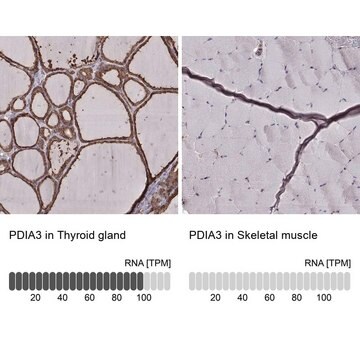
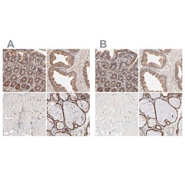
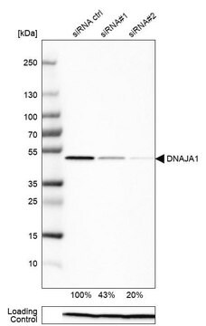
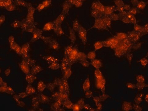
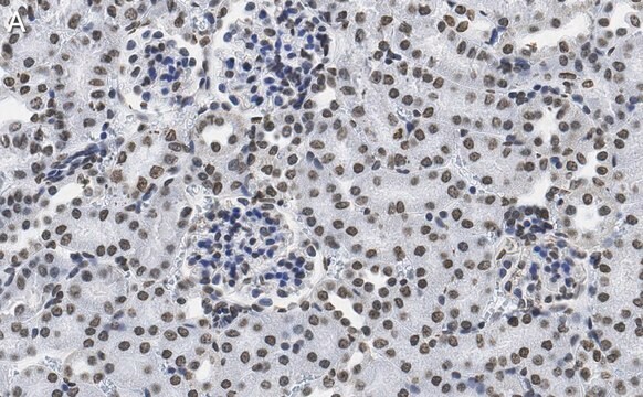
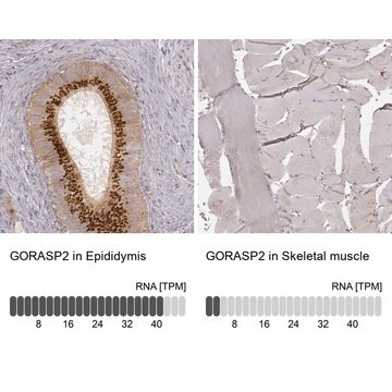
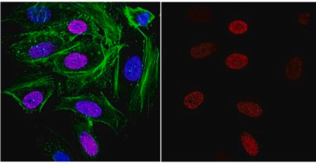
Anti-PDIA3 antibody produced in rabbit, a Prestige Antibody, is developed and validated by the Human Protein Atlas (HPA) project . Each antibody is tested by immunohistochemistry against hundreds of normal and disease tissues. These images can be viewed on the Human Protein Atlas (HPA) site by clicking on the Image Gallery link. The antibodies are also tested using immunofluorescence and western blotting. To view these protocols and other useful information about Prestige Antibodies and the HPA, visit sigma.com/prestige.
Applications in which this antibody has been used successfully, and the associated peer-reviewed papers, are given below.
Immunohistochemistry (1 paper)
Western Blotting (1 paper)

Biochem/physiol Actions

PDIA3 (protein disulfide isomerase family A, member 3) is believed to be a membrane receptor for 1,25(OH)2D3 that stimulates rapid membrane responses through the vitamin D receptor. In the presence of Pdia3, 1,25(OH)2D3 stimulates prostaglandin E2, protein kinase C and other membrane signaling pathways during osteoblast maturation. It stimulates cellular response to chemotherapy-induced DNA damage. The protein functions as a disulfide isomerase protein that associates with lectin chaperones calreticulin and calnexin to mediate folding of glycoproteins. It is found to be secreted in the early stages of renal fibrosis.

Features and Benefits

Prestige Antibodies® are highly characterized and extensively validated antibodies with the added benefit of all available characterization data for each target being accessible via the Human Protein Atlas portal linked just below the product name at the top of this page. The uniqueness and low cross-reactivity of the Prestige Antibodies® to other proteins are due to a thorough selection of antigen regions, affinity purification, and stringent selection. Prestige antigen controls are available for every corresponding Prestige Antibody and can be found in the linkage section.

Every Prestige Antibody is tested in the following ways:
  • IHC tissue array of 44 normal human tissues and 20 of the most common cancer type tissues.
  • Protein array of 364 human recombinant protein fragments.

Linkage

Corresponding Antigen APREST86567

Physical form

Solution in phosphate-buffered saline, pH 7.2, containing 40% glycerol and 0.02% sodium azide

Legal Information

Prestige Antibodies is a registered trademark of Merck KGaA, Darmstadt, Germany

Disclaimer

Unless otherwise stated in our catalog or other company documentation accompanying the product(s), our products are intended for research use only and are not to be used for any other purpose, which includes but is not limited to, unauthorized commercial uses, in vitro diagnostic uses, ex vivo or in vivo therapeutic uses or any type of consumption or application to humans or animals.

Not finding the right product?  

Try our Product Selector Tool.

Storage Class

10 - Combustible liquids

wgk_germany

WGK 1

flash_point_f

Not applicable

flash_point_c

Not applicable

ppe

Eyeshields, Gloves, multi-purpose combination respirator cartridge (US)


Choose from one of the most recent versions:

Certificates of Analysis (COA)

Lot/Batch Number

Don't see the Right Version?

If you require a particular version, you can look up a specific certificate by the Lot or Batch number.

Already Own This Product?

Find documentation for the products that you have recently purchased in the Document Library.

Visit the Document Library

Natalia F Krynetskaia et al.
Molecular cancer therapeutics, 8(4), 864-872 (2009-04-18)
The identification of new molecular components of the DNA damage signaling cascade opens novel avenues to enhance the efficacy of chemotherapeutic drugs. High-mobility group protein 1 (HMGB1) is a DNA damage sensor responsive to the incorporation of nonnatural nucleosides into
Hassan Dihazi et al.
Journal of cell science, 126(Pt 16), 3649-3663 (2013-06-20)
Renal fibrosis is characterized by excessive accumulation of extracellular matrix (ECM), which compromises organ function by replacing normal organ tissue. The molecular mechanisms leading to renal fibrosis are not fully understood. Here we demonstrated that TGFβ1, AGT or PDGF stimulation
Annika Möller-Kerutt et al.
Life science alliance, 6(3) (2022-12-23)
Crumbs2 (CRB2) is a central component of the renal filtration barrier and part of the slit diaphragm, a unique cell contact formed by glomerular podocytes. Some CRB2 variants cause recessive inherited forms of steroid-resistant nephrotic syndrome. However, the disease-causing potential
Huixing Feng et al.
PloS one, 9(4), e95621-e95621 (2014-04-26)
Proteomics study was performed to investigate the specific protein expression profiles of HepG2 cells transfected with mutant HBV compared with wildtype HBV genome, aiming to identify the specific functions of SH3 binding domain (proline rich region) located in HBx. In
Jiaxuan Chen et al.
The Journal of biological chemistry, 285(47), 37041-37050 (2010-09-17)
Protein-disulfide isomerase-associated 3 (Pdia3) is a multifunctional protein hypothesized to be a membrane receptor for 1,25(OH)(2)D(3). In intestinal epithelium and chondrocytes, 1,25(OH)(2)D(3) stimulates rapid membrane responses that are different from genomic effects via the vitamin D receptor (VDR). In this

Our team of scientists has experience in all areas of research including Life Science, Material Science, Chemical Synthesis, Chromatography, Analytical and many others.

Contact Technical Service