Skip to Content
MilliporeSigma
All Photos(1)

Key Documents

A6792

Sigma-Aldrich

Anti-Dog IgG (whole molecule)−Peroxidase antibody produced in rabbit

affinity isolated antibody, buffered aqueous solution

Synonym(s):

Rabbit Anti-Dog IgG (whole molecule)−HRP

Sign Into View Organizational & Contract Pricing


About This Item

MDL number:
UNSPSC Code:
12352203
NACRES:
NA.46

biological source

rabbit

conjugate

peroxidase conjugate

antibody form

affinity isolated antibody

antibody product type

secondary antibodies

clone

polyclonal

form

buffered aqueous solution

technique(s)

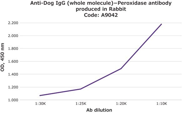
direct ELISA: 1:10,000

shipped in

dry ice

storage temp.

−20°C

target post-translational modification

unmodified

Looking for similar products? Visit Product Comparison Guide

Related Categories

General description

IgG (immunoglobulin G) antibody subtype is the most abundant of serum immunoglobulins of the immune system. It is secreted by B cells and is found in blood and extracellular fluids. Mammalian IgG has four subclasses- IgG1, IgG2a, IgG2b and IgG2c.
Specificity of the anti-dog IgG antiserum is determined by immunoelectrophoresis, prior to conjugation, versus normal dog serum and dog IgG.

Application

Anti-Dog IgG (whole molecule) Peroxidase antibody produced in rabbit has been used:
  • for the detection of antibodies against noro viruses, by ELISA (enzyme linked immunosorbent asay) at 1:2000 for 1 hour at 37°C
  • in IgG (Immunoglobulin G) staining method
  • in ELISA to detect the incorporation of RVGTM (a rabies virus G protein variant)

Biochem/physiol Actions

IgG (Immunoglobulin G) antibody provides protection from infections caused by bacteria, fungi and viruses. Maternal IgG is transferred to fetus through the placenta that is vital for immune defense of the neonate against infections. Anti-Dog IgG antibody is a secondary antibody. Second antibodies (secondary antibodies) are raised against primary antibodies, which bind to specific antigens to create antigen antibody complexes.

Physical form

Solution in 0.01 M phosphate buffered saline, pH 7.4 containing 1% bovine serum albumin and 0.05% MIT

Preparation Note

Prepared using the periodate method described by Wilson, M.B., and Nakane, P.K., in "Immunofluorescence and Related Staining Techniques," Elsevier/North Holland Biomedical Press, Amsterdam, p215 (1978).

Legal Information

Ampliflu is a trademark of Sigma-Aldrich Co. LLC

Disclaimer

Unless otherwise stated in our catalog or other company documentation accompanying the product(s), our products are intended for research use only and are not to be used for any other purpose, which includes but is not limited to, unauthorized commercial uses, in vitro diagnostic uses, ex vivo or in vivo therapeutic uses or any type of consumption or application to humans or animals.

Not finding the right product?  

Try our Product Selector Tool.

pictograms

Exclamation mark

signalword

Warning

hcodes

Hazard Classifications

Skin Sens. 1

Storage Class

12 - Non Combustible Liquids

wgk_germany

WGK 2

flash_point_f

Not applicable

flash_point_c

Not applicable


Certificates of Analysis (COA)

Search for Certificates of Analysis (COA) by entering the products Lot/Batch Number. Lot and Batch Numbers can be found on a product’s label following the words ‘Lot’ or ‘Batch’.

Already Own This Product?

Find documentation for the products that you have recently purchased in the Document Library.

Visit the Document Library

I F G de Amorim et al.
Veterinary parasitology, 173(1-2), 55-63 (2010-07-20)
Dogs represent the major reservoir of Leishmaniao chagasi and vaccination against the canine disease is a potential control strategy. However, seroconversion occurs post-vaccine and hence, there is need to discriminate between the former group and naturally infected dogs. The present
Maria M Figueiredo et al.
BMC immunology, 14, 22-22 (2013-05-15)
Infection with parasite protozoa is a long-term health issue in tropical and subtropical regions throughout the world. The Toll-like receptor (TLR) signaling pathway is one of the first-responding defense systems against Leishmania. The aim of this study was to investigate
Chimeric rabies glycoprotein with a transmembrane domain and cytoplasmic tail from Newcastle disease virus fusion protein incorporates into the Newcastle disease virion at reduced levels
Yu GM, et al.
Journal of Veterinary Science, 18(S1), 351-359 (2017)
Vivian Tamietti Martins et al.
PloS one, 10(9), e0137683-e0137683 (2015-09-15)
In the present study, two Leishmania infantum hypothetical proteins present in the amastigote stage, LiHyp1 and LiHyp6, were combined with a promastigote protein, IgE-dependent histamine-releasing factor (HRF); to compose a polyproteins vaccine to be evaluated against L. infantum infection. Also
Lukas Uhrik et al.
The Biochemical journal, 478(1), 99-120 (2020-12-08)
A comparative canine-human therapeutics model is being developed in B-cell lymphoma through the generation of a hybridoma cell that produces a murine monoclonal antibody specific for canine CD20. The hybridoma cell produces two light chains, light chain-3, and light chain-7.

Our team of scientists has experience in all areas of research including Life Science, Material Science, Chemical Synthesis, Chromatography, Analytical and many others.

Contact Technical Service