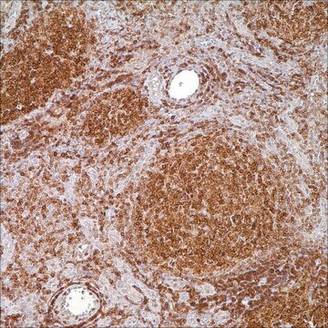
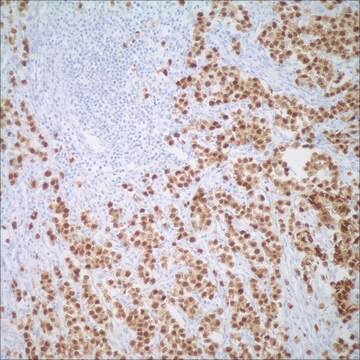
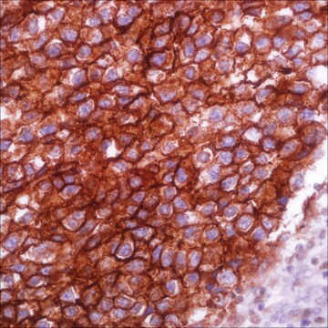
310M-2
Parathyroid Hormone (PTH) (MRQ-31) Mouse Monoclonal Antibody
About This Item
Recommended Products
biological source
mouse
Quality Level
100
500
conjugate
unconjugated
antibody form
culture supernatant
antibody product type
primary antibodies
clone
MRQ-31, monoclonal
description
For In Vitro Diagnostic Use in Select Regions (See Chart)
form
buffered aqueous solution
species reactivity
human
packaging
vial of 0.1 mL concentrate (310M-24)
vial of 0.5 mL concentrate (310M-25)
bottle of 1.0 mL predilute (310M-27)
vial of 1.0 mL concentrate (310M-26)
bottle of 7.0 mL predilute (310M-28)
manufacturer/tradename
Cell Marque™
technique(s)
immunohistochemistry (formalin-fixed, paraffin-embedded sections): 1:100-1:500
isotype
IgG2a
control
parathyroid tissue
shipped in
wet ice
storage temp.
2-8°C
visualization
cytoplasmic
Gene Information
human ... PTH(5741)
General description
Surgical pathologists are familiar with the ability of parathyroid proliferations to assume a variety of histological guises, posing difficulty to categorize any given lesion as hyperplastic, adenomatous or carcinomatous in nature (Wick et al, 1997). This is usually resolved with macroscopic appearance of the remaining parathyroid glands as assessed by the surgeon. The role of the surgical pathologist is to identify the lesion as parathyroid in nature and to assess whether it is normocellular or hypercellular. Although easily accomplished in the majority of instances, rare examples of parathyroid hyperplasia/adenoma showing a follicular/trabecular arrangement may cause concern over the alternative diagnosis of a thyroid adenoma. This becomes more pertinent when the parathyroid lesion abuts into the thyroid gland or lies within the thyroid capsule. Immunostaining for thyroglobulin and parathyroid hormone (PTH) is especially useful to resolve the problem (Permanetter et al, 1983). Nevertheless, caution should be exercised since parathyroid cells often discharge their hormonal product almost as soon as it is packaged in the cytoplasm, resulting in false-negative anti-PTH immunostaining, although the cells are biologically synthetic (Wick et al, 1997)
Anti-PTH antibody is also useful to distinguish parathyroid hyperplasia/neoplasms from thyroid and metastatic neoplasms (Wick et al, 1997); although the pathologist is typically aware of the preoperative hypercalcemic status. Occasionally when the surgeon does not supply this information PTH immunohistochemistry is essential. Even more problematic, are situations in which clear cell parathyroid carcinomas are nonsecretory without an abnormality in mineral metabolism (Aldinger et al, 1982). In such situations, metastatic renal cell carcinoma or metastatic clear cell carcinoma of the lung is evident warranting PTH immunohistochemistry to arrive at the correct diagnosis (Wick et al, 1997). The other instance in which anti-PTH antibodies are useful is in the consideration of parathyroid carcinomas located primarily in the anterior mediastinum (intrathymically). In this situation distinction from primary thymic metastatic carcinomas, non-Hodgkin′s lymphoma and germ cell tumors is necessary (Murphy et al, 1986).
The diagnosis of the majority of parathyroid proliferation may be accomplished with an adequate history, biochemistry profile, and histomorphological assessment; however, rare instances in which the tumors have an abnormal location, clear cell morphology, or a non-secretory may result in erroneous diagnoses, warranting anti-PTH immunohistochemistry.
Quality
 IVD |  IVD |  IVD |  RUO |
Linkage
Physical form
Preparation Note
Other Notes
Legal Information
Not finding the right product?
Try our Product Selector Tool.
Storage Class
12 - Non Combustible Liquids
wgk_germany
WGK 2
flash_point_f
Not applicable
flash_point_c
Not applicable
Certificates of Analysis (COA)
Search for Certificates of Analysis (COA) by entering the products Lot/Batch Number. Lot and Batch Numbers can be found on a product’s label following the words ‘Lot’ or ‘Batch’.
Already Own This Product?
Find documentation for the products that you have recently purchased in the Document Library.
Our team of scientists has experience in all areas of research including Life Science, Material Science, Chemical Synthesis, Chromatography, Analytical and many others.
Contact Technical Service