Skip to Content
MilliporeSigma
All Photos(1)

Key Documents

229R-1

Sigma-Aldrich

Calcitonin (SP17) Rabbit Monoclonal Antibody

Sign Into View Organizational & Contract Pricing


About This Item

UNSPSC Code:
12352203
NACRES:
NA.41

biological source

rabbit

Quality Level

100
500

conjugate

unconjugated

antibody form

culture supernatant

antibody product type

primary antibodies

clone

SP17, monoclonal

description

For In Vitro Diagnostic Use in Select Regions (See Chart)

form

buffered aqueous solution

species reactivity

human

packaging

vial of 0.1 mL concentrate (229R-14)
vial of 0.5 mL concentrate (229R-15)
bottle of 1.0 mL predilute (229R-17)
vial of 1.0 mL concentrate (229R-16)
bottle of 7.0 mL predilute (229R-18)

manufacturer/tradename

Cell Marque

technique(s)

immunohistochemistry (formalin-fixed, paraffin-embedded sections): 1:100-1:500

isotype

IgG

control

medullary carcinoma of thyroid

shipped in

wet ice

storage temp.

2-8°C

visualization

cytoplasmic

General description

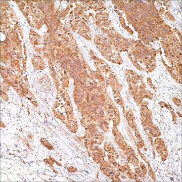
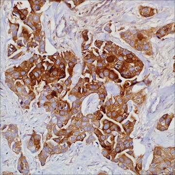
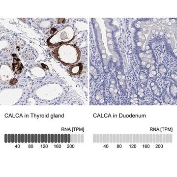
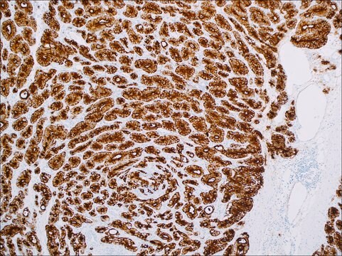
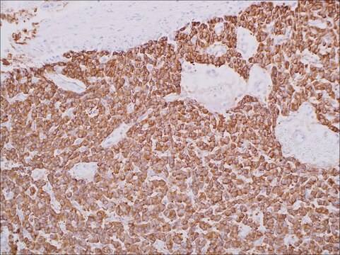
Calcitonin (thyrocalcitonin) is a 32 amino acid linear polypeptide hormone that is involved in regulating calcium metabolism. It is primarily produced by the parafollicular cells (also known as C cells) of the thyroid gland, though some neuroendocrine cells in other tissues, such as the respiratory and digestive tracts, can also synthesize calcitonin. Anti-calcitonin immunohistochemistry is an effective way for demonstrating the parafollicular C cells in the thyroid3,4 and the tumors arising from them, including C cell hyperplasia of the thyroid and medullary thyroid carcinoma, a neuroendocrine tumor that originates from C cells.
Immunohistochemical staining with anti-calcitonin antibody has proven to be an effective way of demonstrating calcitonin-producing cells in the thyroid. C-cell hyperplasia and medullary thyroid carcinomas stain positive for calcitonin. Studies of calcitonin have resulted in the identification of a wide spectrum of C-cell proliferative abnormalities. citationcitationcitationcitationcitation

Quality


IVD

IVD

IVD

RUO

Linkage

Calcitonin Positive Control Slides, Product No. 229S, are available for immunohistochemistry (formalin-fixed, paraffin-embedded sections).

Physical form

Solution in Tris Buffer, pH 7.3-7.7, with 1% BSA and <0.1% Sodium Azide

Preparation Note

Download the IFU specific to your product lot and formatNote: This requires a keycode which can be found on your packaging or product label.

Other Notes

For Technical Service please contact: 800-665-7284 or email: service@cellmarque.com

Legal Information

Cell Marque is a trademark of Merck KGaA, Darmstadt, Germany

Not finding the right product?  

Try our Product Selector Tool.


Certificates of Analysis (COA)

Search for Certificates of Analysis (COA) by entering the products Lot/Batch Number. Lot and Batch Numbers can be found on a product’s label following the words ‘Lot’ or ‘Batch’.

Already Own This Product?

Find documentation for the products that you have recently purchased in the Document Library.

Visit the Document Library

Plasma-immunoreactive-calcitonin in patients with non-thyroid tumours.
R C Coombes et al.
Lancet (London, England), 1(7866), 1080-1083 (1974-06-01)
Evidence for calcitonin--a new hormone from the parathyroid that lowers blood calcium.
D H COPP et al.
Endocrinology, 70, 638-649 (1962-05-01)
Y Dayal et al.
Cancer, 43(4), 1331-1338 (1979-04-01)
A nonfunctioning strumal carcinoid arising in a 49-year-old woman was studied by histochemical and immunocytochemical techniques. All tumor cells, irrespective of their architectural arrangement, showed properties of neuroendocrine-programmed cells, without any evidence of thyroid follicular cell differentiation. Foci of calcitonin-producing
Calcitonin and carcinoembryonic antigen as tumor markers in medullary thyroid carcinoma.
R A DeLellis et al.
American journal of clinical pathology, 70(4), 587-594 (1978-10-01)
Y Kameda et al.
Cell and tissue research, 206(3), 403-415 (1980-01-01)
The development of C-cell complexes was investigated in dog fetuses by an immunoperoxidase method with three specific antisera: anti-calcitonin, anti-C-thyroglobulin (C-Tg), and anti-19S thyroglobulin. Ultimobranchial bodies joined with the thyroid anlage and then dispersed into the parenchyma to form large

Our team of scientists has experience in all areas of research including Life Science, Material Science, Chemical Synthesis, Chromatography, Analytical and many others.

Contact Technical Service