Recommended Products
biological source
bacterial (Nocardia otitidis-caviarum)
Quality Level
form
solution
packaging
pkg of 1,000 U (11014714001 [10 U/μl])
pkg of 1,000 U (11037668001 [40 U/μl])
pkg of 200 U (11014706001 [10 U/μl])
manufacturer/tradename
Roche
parameter
37 °C optimum reaction temp.
technique(s)
PCR: suitable
storage temp.
−20°C
Related Categories
General description
Not I belongs to the class of "rare-cutter" enzymes. It is one of the two known enzymes that recognize an octameric sequence comprised solely of G and C residues.
Contents:
- Not I
- SuRE/Cut Buffer H (10x)
Specificity
GCGG*C*CG °C
Restriction site: GC↓GG*C*CG °C
GC↓GG*C*CG °C
Heat inactivation: Not I can be heat inactivated by incubation at 65 °C for 15 minutes (up to 100 U/μg DNA).
Quality
1 μg Ad2 DNA is incubated for 16 hours in 50 μl SuRE/Cut Buffer H with an excess of Not I. The number of enzyme units which do not change the enzyme-specific pattern is stated in the certificate of analysis.
Absence of exonuclease activity
Approximately 5 μg [3H] labeled calf thymus DNA are incubated with 3 μl Not I for 4 hours at +37°C in a total volume of 100 μl 50 mM Tris-HCl, 10 mM MgCl2, 1 mM Dithioerythritol, pH approximately 7.5. Under these conditions, no release of radioactivity is detectable, as stated in the certificate of analysis.
Specifications
Prokaryotic genomic DNA: Not I fragments are between 20 and 1,000 kb, depending on the GC content.
Yeast genomic DNA: Not I fragments are, on average, 200 kb.
Mammalian genomic DNA: Not I fragments are approximately 1,000 kb.
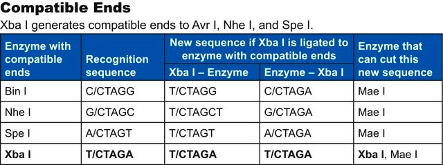
Compatible ends
Not I ends are compatible with ends generated by Eae I and EclX I (Xma III).
Isoschizomers
The enzyme has no known isoschizomers.
Methylation sensitivity
Not I is inhibited by the presence of 5-methylcytosine at the sites indicated (*) on the recognition sequence. However, the presence of 5-methylcytosine in the 5′-C position (°) is not inhibiting.
Relative activity in complete PCR mix
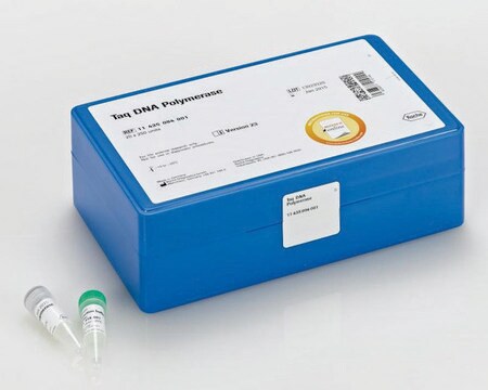
Relative activity in PCR mix (Taq DNA Polymerase buffer) is 0%. The PCR mix contained λDNA, primers, 10 mM Tris-HCl (pH 8.3, +20°C), 50 mM KCl, 1.5 mM MgCl2, 200 μM dNTPs, 2.5 U Taq DNA polymerase. The mix was subjected to 25 amplification cycles. After addition of 100 mM NaCl to the RE digest in the PCR mix, the activity of Not I still remains very low with below 5%.
Incubation temperature
+37°C
PFGE tested
Not I has been tested in Pulsed-Field Gel Electrophoresis (on bacterial chromosomes). For cleavage of genomic DNA (E. coli C 600) embedded in agarose for PFGE analysis, we recommend using 10 U of enzyme/μg DNA and 4 hour incubation time.
Ligation and recutting assay
Not I fragments obtained by complete digestion of 1 μg Ad2 DNA are ligated with 1 U T4 DNA Ligase in a volume of 10 μl by incubation for 16 hours at +4°C in 66 mM Tris-HCl, 5 mM MgCl2, 5 mM Dithiothreitol, 1 mM ATP, pH 7.5 (at +20°C) resulting in >80% recovery of Ad2 DNA.
Subsequent re-cutting with Not I yields >90% of the typical pattern of Ad2 × Not I fragments.
DNA Profile
- λ: 0
- φX174: 0
- Ad2: 7
- M13mp7: 0
- pBR322: 0
- pBR328: 0
- pUC18: 0
- SV40: 0
Unit Definition
Analysis Note
The buffer in bold is recommended for optimal activity
- A: 10-25%
- B: 50-75%
- H: 100%
- L: 0-10%
- M: 25-50%
Other Notes
Kit Components Only
- Enzyme Solution
- SuRE/Cut Buffer H 10x concentrated
Storage Class
12 - Non Combustible Liquids
wgk_germany
WGK 1
flash_point_f
does not flash
flash_point_c
does not flash
Certificates of Analysis (COA)
Search for Certificates of Analysis (COA) by entering the products Lot/Batch Number. Lot and Batch Numbers can be found on a product’s label following the words ‘Lot’ or ‘Batch’.
Already Own This Product?
Find documentation for the products that you have recently purchased in the Document Library.
Articles
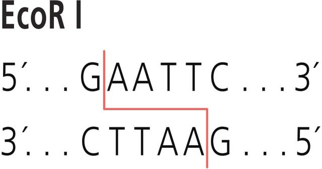
The term “Restriction enzyme” originated from the studies of Enterobacteria phage λ (lambda phage) in the laboratories of Werner Arber and Matthew Meselson.
Related Content
Restriction endonucleases popularly referred to as restriction enzymes, are ubiquitously present in prokaryotes. The function of restriction endonucleases is mainly protection against foreign genetic material especially against bacteriophage DNA.
Our team of scientists has experience in all areas of research including Life Science, Material Science, Chemical Synthesis, Chromatography, Analytical and many others.
Contact Technical Service