Skip to Content
MilliporeSigma
All Photos(2)

Key Documents

MABS1261

Sigma-Aldrich

Anti-FRA-2 Antibody, clone REY146C

clone REY146C, from rat

Synonym(s):

Fos-related antigen 2, FRA-2

Sign Into View Organizational & Contract Pricing


About This Item

UNSPSC Code:
12352203
eCl@ss:
32160702
NACRES:
NA.41
clone:
REY146C, monoclonal
application:
ChIP
IF
IHC
IP
WB
species reactivity:
mouse
technique(s):
ChIP: suitable
immunofluorescence: suitable
immunohistochemistry: suitable (paraffin)
immunoprecipitation (IP): suitable
western blot: suitable
citations:
4

biological source

rat

Quality Level

antibody form

purified immunoglobulin

antibody product type

primary antibodies

clone

REY146C, monoclonal

species reactivity

mouse

technique(s)

ChIP: suitable
immunofluorescence: suitable
immunohistochemistry: suitable (paraffin)
immunoprecipitation (IP): suitable
western blot: suitable

isotype

IgG2aκ

NCBI accession no.

UniProt accession no.

shipped in

wet ice

target post-translational modification

unmodified

Gene Information

mouse ... Fosl2(14284)

General description

Fos-related antigen 2 (UniProt P47930; also known as FRA-2) is encoded by the Fosl2 (also known as Fra-2, Fra2) gene (Gene ID 14284) in murine species. FRA-2 belongs to the family of AP-1 proteins consisting of the Fos (c-Fos, FosB, Fra-1, and Fra-2) and Jun (c-Jun, JunB, and JunD) members that act as sequence-specific homodimeric and heterodimeric transcription factors activated by mitogen-activated protein (MAP) kinases, including JNK, p38, and ERK. The majority of terminal differentiation proteins are encoded by genes located within the epidermal differentiation complex (EDC) of basal keratinocytes. EDC genes are transcriptionally repressed by the chromatin-remodeling polycomb repressor complex (PRC) via histone H3 trimethylation on Lys27 (H3K27me3) and transcribed by the AP-1 transcription factor activity in the absence of H3K27me3 modification. Fra-2 specifically is shown to regulate the expression of EDC genes by direct promoter binding upon activation by ERK1/2-dependent phosphorylation. Deregulated expression of AP-1 proteins causes skin diseases and epithelial cancers, such as psoriasis and squamous cell carcinomas (SCCs). Elevated levels of FRA2 can serve as an indicator of caerulein-induced inflammation in pancreas. (Ref. Cobo, I et al (2018). Nature 554(7693); 533-537).

Specificity

Clone REY146C reacts with murine FRA-2 (mFRA-2), but not mFRA-1 or mFosB. Expected to react with both spliced isoforms of mFRA-2 (Uniprot P47930-1 and P47930-2).

Immunogen

Epitope: C-terminal half.
His-tagged recombinant protein corresponding to the C-terminal half of mouse FRA-2.

Application

Anti-FRA-2 Antibody, clone REY146C is an antibody against FRA-2 for use in Western Blotting, Immunofluorescence, Immunoprecipitation, Chromatin Immunoprecipitation (ChIP), Immunohistochemistry (Paraffin).
Immunofluorescence Analysis: A representative lot detected FRA-2 in all epidermal layers with the highest expression in terminally differentiated keratinocytes in OCT-embedded frozen back skin sections from E17.5 mouse embryos (Wurm, S., et al. (2014). Genes Dev. 15;29(2):144-156).
Western Blotting Analysis: A representative lot detected a FRA-2 upregulation in cultured mouse keratinocytes (mKCs) upon Ca2+-induced differentiation. ERK1/2 inhibitor FR180204 (Cat. No. 328007) reduced Fra-2 level in mKCs upon Ca2+ treatment (Wurm, S., et al. (2014). Genes Dev. 15;29(2):144-156).
Immunoprecipitation Analysis: A representative lot co-immunoprecipitated Ezh2 with Fra-2 from lysates of cultured mouse keratinocytes (mKCs) prior to, but not after, Ca2+-induced differentiation (Wurm, S., et al. (2014). Genes Dev. 15;29(2):144-156).
Chromatin Immunoprecipitation Analysis: A representative lot detected FRA-2 association with EDC promoters at conserved AP-1 consensus sites in cultured mouse keratinocytes (mKCs) under both basal and differentiation conditions (Wurm, S., et al. (2014). Genes Dev. 15;29(2):144-156).
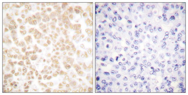
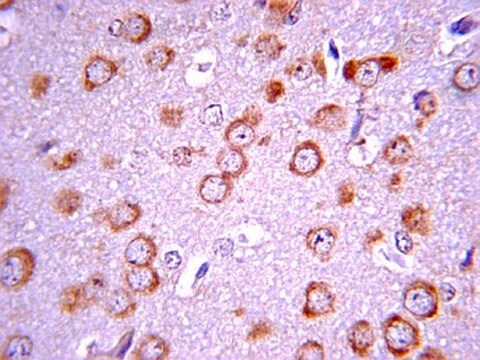
Immunohistochemistry Analysis: Prior to antibody purification, the culture supernatant detected FRA-2 immunoreactivity in paraffin-embedded skin sections from wild-type, but not Fosl2-knockout mice (Courtesy of Dr. Erwin Wagner, Spanish National Cancer Research Centre (CNIO), Madrid, Spain).
Immunocytochemistry Analysis: Prior to antibody purification, the culture supernatant detected exogenously expressed murine FRA-2 (mFRA-2), but not mFRA-1 or mFosB in transfected HEK293T cells (Courtesy of Dr. Erwin Wagner, Spanish National Cancer Research Centre (CNIO), Madrid, Spain).
Research Category
Signaling
Research Sub Category
Transcription Factors

Quality

Evaluated by Western Blotting in MEF1 cell lysate.

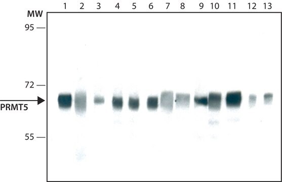
Western Blotting Analysis: 0.5 µg/mL of this antibody detected FRA-2 in 10 µg of MEF1 cell lysate.

Target description

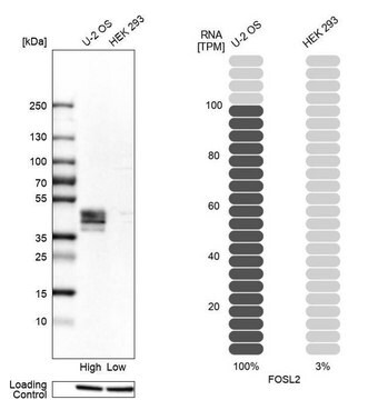
~41 and 38 kDa observed.

Physical form

Format: Purified
Protein G Purified
Purified rat monoclonal IgG2aκ antibody in buffer containing 0.1 M Tris-Glycine (pH 7.4), 150 mM NaCl with 0.05% sodium azide.

Storage and Stability

Stable for 1 year at 2-8°C from date of receipt.

Other Notes

Concentration: Please refer to lot specific datasheet.

Disclaimer

Unless otherwise stated in our catalog or other company documentation accompanying the product(s), our products are intended for research use only and are not to be used for any other purpose, which includes but is not limited to, unauthorized commercial uses, in vitro diagnostic uses, ex vivo or in vivo therapeutic uses or any type of consumption or application to humans or animals.

Not finding the right product?  

Try our Product Selector Tool.

Storage Class

12 - Non Combustible Liquids

wgk_germany

WGK 1

flash_point_f

Not applicable

flash_point_c

Not applicable


Certificates of Analysis (COA)

Search for Certificates of Analysis (COA) by entering the products Lot/Batch Number. Lot and Batch Numbers can be found on a product’s label following the words ‘Lot’ or ‘Batch’.

Already Own This Product?

Find documentation for the products that you have recently purchased in the Document Library.

Visit the Document Library

Mike R Wilson et al.
Human reproduction (Oxford, England), 35(1), 58-69 (2019-12-31)
Is it possible to establish a genetically engineered mouse model (GEMM) of endometriosis that mimics the natural spread of invasive endometrium? Endometriosis occurs in an ARID1A (AT-rich interactive domain-containing protein 1A) and PIK3CA (phosphatidylinositol-4,5-bisphosphate 3-kinase, catalytic subunit alpha) mutant GEMM

Our team of scientists has experience in all areas of research including Life Science, Material Science, Chemical Synthesis, Chromatography, Analytical and many others.

Contact Technical Service