
MAB3418
Anti-MAP2 Antibody
CHEMICON®, mouse monoclonal, AP20
Synonym(s):
Microtubule Associated Protein, MAP2A and 2B
About This Item
Recommended Products


Product Name
Anti-MAP2 Antibody, clone AP20, clone AP20, Chemicon®, from mouse
biological source
mouse
Quality Level
antibody form
purified immunoglobulin
antibody product type
primary antibodies
clone
AP20, monoclonal
species reactivity
bovine, mouse, rat, chicken, quail, Xenopus, human
packaging
antibody small pack of 25 μg
manufacturer/tradename
Chemicon®
technique(s)
immunohistochemistry: suitable
western blot: suitable
isotype
IgG
NCBI accession no.
UniProt accession no.
shipped in
ambient
storage temp.
2-8°C
target post-translational modification
unmodified
Gene Information
chicken ... Map2(424001)
frog ... Map2(780043)
human ... MAP2(4133)
mouse ... Map2(17756) , Map2(281294)
rat ... Map2(25595)
General description
Specificity
Immunogen
Application
Neuroscience
Neuronal & Glial Markers
60 ng/mL. In SDS-PAGE, MAP-2 from adult rat migrates as a closely associated doublet having a molecular weight of approximately 280-300 kD representing the MAP-2A and -2B isoforms but not the 70 kDa MAP-2C isoform. However, early in brain development (postnatal day 10 in rats), MAP-2 migrates as a single band that is identical to the lower molecular weight band of the adult MAP-2 doublet (MAP-2b). Later in development (postnatal days 17-18), the mobility of MAP-2 changes to the adult doublet form. (In the spinal cord, conversion to the adult form occurs earlier). Users should run 4%-20% SDS-PAGE gradient gels.
Immunohistochemistry:
5 µg/mL (4% PFA fixed frozen sections).
Immunohistochemistry:
5 µg/mL of a previous lot was used. Anti-MAP-2 can be used to stain tissue (brain or spinal cord) fixed with paraformaldehyde.
Optimal working concentrations must be determined by the end user.
Quality
Western Blot Analysis:
1:1000 dilution of this lot detected MAP2 on 10 μg of Rat Brain lysate.
Target description
Physical form
Storage and Stability
During shipment, small volumes of product will occasionally become entrapped in the seal of the product vial. For products with volumes of 200 µL or less, we recommend gently tapping the vial on a hard surface or briefly centrifuging the vial in a tabletop centrifuge to dislodge any liquid in the container’s cap.
Analysis Note
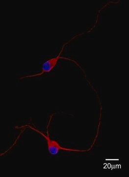
Brain, Neuronal culture, Human glioblastoma T98G cells.
Other Notes
Legal Information
Disclaimer
Not finding the right product?
Try our Product Selector Tool.
recommended
Storage Class
12 - Non Combustible Liquids
wgk_germany
WGK 1
flash_point_f
Not applicable
flash_point_c
Not applicable
Certificates of Analysis (COA)
Search for Certificates of Analysis (COA) by entering the products Lot/Batch Number. Lot and Batch Numbers can be found on a product’s label following the words ‘Lot’ or ‘Batch’.
Already Own This Product?
Find documentation for the products that you have recently purchased in the Document Library.
Customers Also Viewed


Articles
Human iPSC neural differentiation media and protocols used to generate neural stem cells, neurons and glial cell types.
Our team of scientists has experience in all areas of research including Life Science, Material Science, Chemical Synthesis, Chromatography, Analytical and many others.
Contact Technical Service