MAB1083
Anti-GFP Antibody, clone 3F8.2
clone 3F8.2, from mouse
Synonym(s):
Green Fluorescent protein
Sign Into View Organizational & Contract Pricing
All Photos(1)
About This Item
UNSPSC Code:
12352203
eCl@ss:
32160702
NACRES:
NA.41
Recommended Products
biological source
mouse
Quality Level
antibody form
purified immunoglobulin
antibody product type
primary antibodies
clone
3F8.2, monoclonal
species reactivity
vertebrates
technique(s)
western blot: suitable
isotype
IgG1κ
UniProt accession no.
shipped in
wet ice
target post-translational modification
unmodified
General description
The Green Fluorescent Protein (GFP) from the jellyfish Aequorea victoria is used as a fluorescent indicator for monitoring gene expression in a variety of cellular systems, including living organisms and fixed tissues. Unlike other bioluminescent reporters, GFP fluoresces in the absence of substrates, cofactors, or other intrinsic or extrinsic proteins. Purified GFP is a 27 kDa monomer consisting of 238 amino acids and emits green light (emission maximum at 509 nm) when excited with blue or UV light.
Specificity
Reacts with most common vertebrate proteins containing the GFP tag.
The antibody reacts with the 28 kDa GFP protein.
Immunogen
Epitope: Full Length
Full length GFP protein.
Application
Detect GFP using this Anti-GFP Antibody, clone 3F8.2 validated for use in WB.
Research Category
Secondary & Control Antibodies
Secondary & Control Antibodies
Research Sub Category
Epitope Tags
Epitope Tags
Western Blot Analysis:
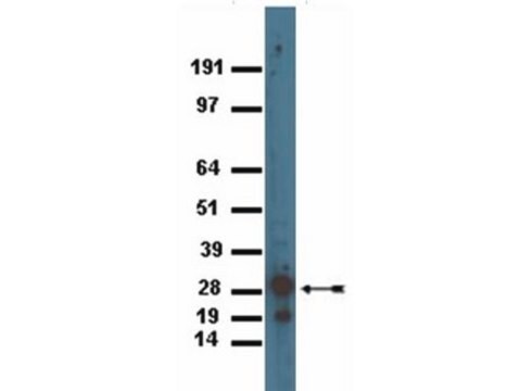
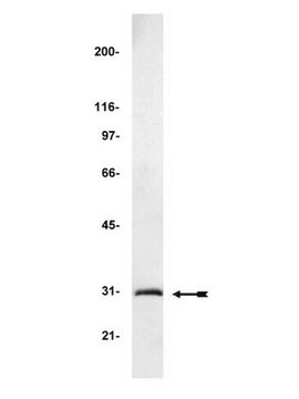
0.125 μg/mL - 1 μg/mL of this antibody detected GFP in 10 ng of GFP recombinant protein (Cat no. 14-392).
0.125 μg/mL - 1 μg/mL of this antibody detected GFP in 10 ng of GFP recombinant protein (Cat no. 14-392).
Quality
Routinely evaluated by Western Blot on GFP recombinant protein.
Target description
28 kDa
Physical form
Format: Purified
Protein G Purified
Purified mouse immunoglobulin in buffer containing PBS containing 0.05% sodium azide.
Storage and Stability
Maintain at 2-8°C in undiluted aliquots for up to 1 year after date of receipt. Avoid repeated freeze/thaw cycles, which may damage IgG and affect product performance.
Analysis Note
Control
Western Blot: GFP recombinant protein
Western Blot: GFP recombinant protein
Other Notes
Concentration: Please refer to the Certificate of Analysis for the lot-specific concentration.
Disclaimer
Unless otherwise stated in our catalog or other company documentation accompanying the product(s), our products are intended for research use only and are not to be used for any other purpose, which includes but is not limited to, unauthorized commercial uses, in vitro diagnostic uses, ex vivo or in vivo therapeutic uses or any type of consumption or application to humans or animals.
Not finding the right product?
Try our Product Selector Tool.
recommended
Storage Class
12 - Non Combustible Liquids
wgk_germany
WGK 1
flash_point_f
Not applicable
flash_point_c
Not applicable
Certificates of Analysis (COA)
Search for Certificates of Analysis (COA) by entering the products Lot/Batch Number. Lot and Batch Numbers can be found on a product’s label following the words ‘Lot’ or ‘Batch’.
Already Own This Product?
Find documentation for the products that you have recently purchased in the Document Library.
Customers Also Viewed
Human cardiovascular progenitor cells develop from a KDR+ embryonic-stem-cell-derived population.
Yang, Lei, et al.
Nature, 453, 524-528 (2008)
Bernadette M DeRussy et al.
Journal of virology, 90(16), 7109-7117 (2016-05-27)
Human cytomegalovirus (HCMV) pUL93 and pUL77 are both essential for virus growth, but their functions in the virus life cycle remain mostly unresolved. Homologs of pUL93 and pUL77 in herpes simplex virus 1 (HSV-1) and pseudorabies virus (PRV) are known
Jake D Howden et al.
BMC biology, 19(1), 130-130 (2021-06-24)
Keratinocytes form the main protective barrier in the skin to separate the underlying tissue from the external environment. In order to maintain this barrier, keratinocytes form robust junctions between neighbouring cells as well as with the underlying extracellular matrix. Cell-cell
Markus S Spurlock et al.
Journal of neurotrauma, 34(11), 1981-1995 (2017-03-03)
Penetrating traumatic brain injury (PTBI) is one of the major cause of death and disability worldwide. Previous studies with penetrating ballistic-like brain injury (PBBI), a PTBI rat model revealed widespread perilesional neurodegeneration, similar to that seen in humans following gunshot
Our team of scientists has experience in all areas of research including Life Science, Material Science, Chemical Synthesis, Chromatography, Analytical and many others.
Contact Technical Service