Skip to Content
MilliporeSigma
All Photos(2)

Key Documents

ABN90P

Sigma-Aldrich

Anti-NeuN purified Antibody

from guinea pig, purified by affinity chromatography

Synonym(s):

NEUronal Nuclei, clone A60, NeuN

Sign Into View Organizational & Contract Pricing


About This Item

UNSPSC Code:
12352203
eCl@ss:
32160702
NACRES:
NA.41

biological source

guinea pig

Quality Level

antibody form

affinity isolated antibody

antibody product type

primary antibodies

clone

polyclonal

purified by

affinity chromatography

species reactivity

human, rat, mouse

technique(s)

immunocytochemistry: suitable
immunohistochemistry: suitable (paraffin)
western blot: suitable

shipped in

wet ice

target post-translational modification

unmodified

Gene Information

General description

ABN90P is a Guinea Pig polyclonal version of the Anti-NeuN, clone A60 (MAB377), a highly characterized and cited mouse monoclonal antibody that specifically recognizes the DNA-binding, neuron-specific protein NeuN, which is present in most CNS and PNS neuronal cell types of all vertebrates tested. NeuN protein distributions are apparently restricted to neuronal nuclei, perikarya and some proximal neuronal processes in both fetal and adult brain although, some neurons fail to be recognized by NeuN at all ages: INL retinal cells, Cajal-Retzius cells, Purkinje cells, inferior olivary and dentate nucleus neurons, and sympathetic ganglion cells are some examples. Immunohistochemically detectable NeuN protein first appears at developmental timepoints that correspond with the withdrawal of the neuron from the cell cycle and/or with the initiation of terminal differentiation of the neuron. Immunoreactivity appears around E9.5 in the mouse neural tube and is extensive throughout the developing nervous system by E12.5. Strong nuclear staining suggests a nuclear regulatory protein function. No difference between protein isolated from purified nuclei and whole brain extract on immunoblots has been found.

Specificity

This antibody recognizes the N-terminus of NeuN.

Immunogen

Epitope: N-terminus
GST-tagged recombinant protein corresponding to the N-terminus of mouse NeuN.

Application

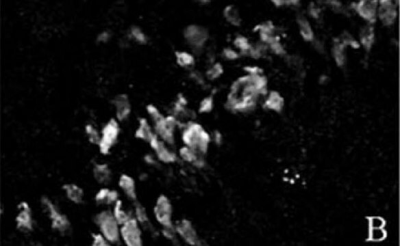
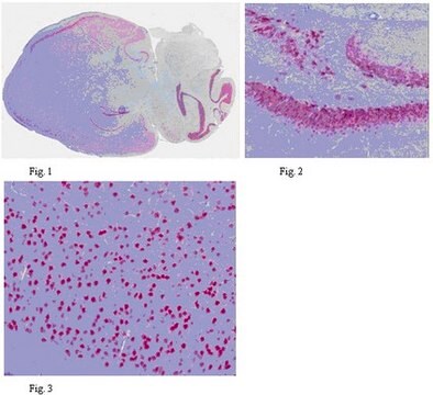
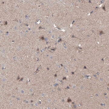
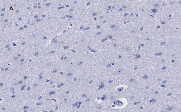
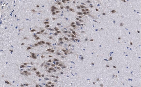
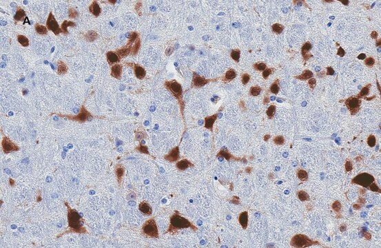
Immunohistochemistry Analysis: A 1:1,000 dilution from a representative lot detected NeuN in mouse frontal cortex tissue.

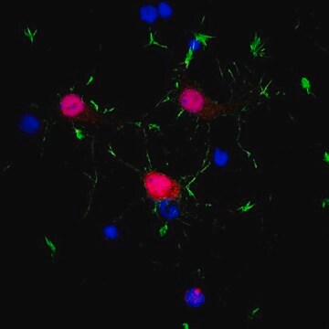
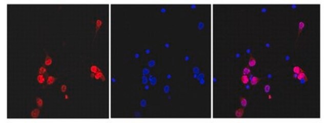
Immunocytochemistry Analysis: A 1:1,000-2,000 dilution from a representative lot detected NeuN in rat E18 cortex cells.
Research Category
Neuroscience
Research Sub Category
Neuronal & Glial Markers
This Anti-NeuN purified Antibody is validated for use in Western Blotting, ICC, IHC(P) for the detection of NeuN purified.

Quality

Evaluated by Western Blot in mouse E16 brain tissue lysate.

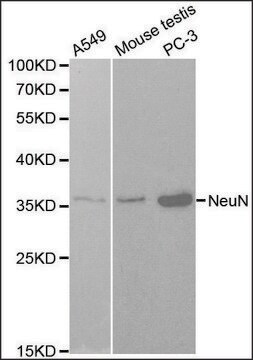
Western Blot Analysis: A 1:1,000 dilution of this antibody detected NeuN on 10 µg of mouse E16 brain tissue lysate.

Target description

~48 kDa observed

Physical form

Antigen Affinity Purified
Purified guinea pig polyclonal in buffer containing 0.1 M Tris-Glycine (pH 7.4), 150 mM NaCl with 0.05% sodium azide.

Storage and Stability

Stable for 1 year at 2-8°C from date of receipt.

Analysis Note

Control
Mouse E16 brain tissue lysate

Other Notes

Concentration: Please refer to the Certificate of Analysis for the lot-specific concentration.

Disclaimer

Unless otherwise stated in our catalog or other company documentation accompanying the product(s), our products are intended for research use only and are not to be used for any other purpose, which includes but is not limited to, unauthorized commercial uses, in vitro diagnostic uses, ex vivo or in vivo therapeutic uses or any type of consumption or application to humans or animals.

Not finding the right product?  

Try our Product Selector Tool.

Storage Class

12 - Non Combustible Liquids

wgk_germany

WGK 1

flash_point_f

Not applicable

flash_point_c

Not applicable


Certificates of Analysis (COA)

Search for Certificates of Analysis (COA) by entering the products Lot/Batch Number. Lot and Batch Numbers can be found on a product’s label following the words ‘Lot’ or ‘Batch’.

Already Own This Product?

Find documentation for the products that you have recently purchased in the Document Library.

Visit the Document Library

Dianne E Lee et al.
Molecular imaging, 13 (2014-09-25)
The dopaminergic system is especially vulnerable to the effects of human immunodeficiency virus (HIV) infection, rendering dopaminergic deficits early surrogate markers of HIV-associated neuropathology. We quantified dopamine D2/3 receptors in young HIV-1 transgenic (Tg) (n  =  6) and age-matched control
Loïc J Chareyron et al.
Proceedings of the National Academy of Sciences of the United States of America, 113(50), 14420-14425 (2016-12-03)
A large population of immature neurons is present in the ventromedial portion of the adult primate amygdala, a region that receives substantial direct projections from the hippocampal formation. Here, we show the effects of neonatal (n = 8) and adult
Kirsty Sawicka et al.
eLife, 8 (2019-12-21)
Loss of the RNA binding protein FMRP causes Fragile X Syndrome (FXS), the most common cause of inherited intellectual disability, yet it is unknown how FMRP function varies across brain regions and cell types and how this contributes to disease
William Schreiber-Stainthorp et al.
Nature communications, 12(1), 2855-2855 (2021-05-19)
Ebola virus (EBOV) causes neurological symptoms yet its effects on the central nervous system (CNS) are not well-described. Here, we longitudinally assess the acute effects of EBOV on the brain, using quantitative MR-relaxometry, 18F-Fluorodeoxyglucose PET and immunohistochemistry in a monkey
Jun Nishiyama et al.
Neuron, 96(4), 755-768 (2017-10-24)
Precise genome editing via homology-directed repair (HDR) in targeted cells, particularly in vivo, provides an invaluable tool for biomedical research. However, HDR has been considered to be largely restricted to dividing cells, making it challenging to apply the technique in postmitotic

Our team of scientists has experience in all areas of research including Life Science, Material Science, Chemical Synthesis, Chromatography, Analytical and many others.

Contact Technical Service