Skip to Content
MilliporeSigma
All Photos(5)

Key Documents

05-661

Sigma-Aldrich

Anti-β-Tubulin Antibody, clone AA2

clone AA2, Upstate®, from mouse

Synonym(s):

Tubulin beta-5 chain, beta 5-tubulin, beta Ib tubulin, beta-4 tubulin, tubulin beta polypeptide, tubulin beta-1 chain, tubulin, beta, tubulin, beta polypeptide

Sign Into View Organizational & Contract Pricing


About This Item

UNSPSC Code:
12352203
eCl@ss:
32160702
NACRES:
NA.41

biological source

mouse

antibody form

purified immunoglobulin

antibody product type

primary antibodies

clone

AA2, monoclonal

species reactivity

bovine, human, mouse, rat

manufacturer/tradename

Upstate®

technique(s)

western blot: suitable

isotype

IgG1

NCBI accession no.

UniProt accession no.

shipped in

dry ice

target post-translational modification

unmodified

Gene Information

human ... TUBB(203068)
mouse ... Tubb3(22152)

General description

Microtubules are constituent parts of the mitotic apparatus, cilia, flagella, and elements of the cytoskeleton. They consist principally of 2 soluble proteins, alpha- and beta-tubulin, each about 55 kDa in size.
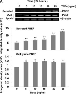
Antibodies against beta Tubulin are useful as loading controls for Western Blotting. However it should be noted that levels of beta Tubulin may not be stable in certain cells. For example, expression of tubulin in adipose tissue is very low (Spiegelman and Farmer, Cell, 1982, 29(1):53-60) and therefore beta Tubulin should not be used as loading control for these tissues.

Specificity

Predicted to have broad species cross-reactivity based on sequence homology.
This antibody recognizes β-Tubulin isotypes I, II, III, IVa, and IVb of ~50 kDa. No cross-reactivity with alpha or gamma tubulin.

Immunogen

Epitope: The epitope has been mapped to amino acids 412-430
Purified bovine brain tubulin. The epitope recognized by this antibody has been mapped to amino acids 412-430.

Application

Research Category
Cell Structure
Research Sub Category
Cytoskeleton
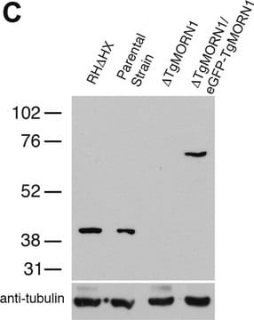
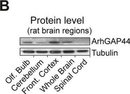
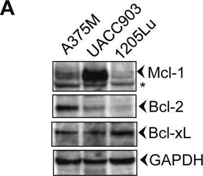
This Anti-β-Tubulin Antibody, clone AA2 is validated for use in WB for the detection of β-Tubulin.

Quality

Routinely evaluated by western blot on RIPA lysates from 3T3/A31, PC-12, Jurkat, or A431 cells.

Western Blot Analysis:
0.1-1.0 µg/mL of this lot detected β-Tubulin in RIPA lysates from 3T3/A31, PC-12, Jurkat, and A431 cells.

Target description

50 kDa

Physical form

Format: Purified
Protein G Purified
Purified mouse monoclonal IgG1 in buffer containing PBS with 0.05% sodium azide and 30% glycerol.

Storage and Stability

Stable for 1 year at -20°C from date of receipt.

Handling Recommendations:
Upon receipt, and prior to removing the cap, centrifuge the vial and gently mix the solution. Aliquot into microcentrifuge tubes and store at -20ºC. Avoid repeated freeze/thaw cycles, which may damage IgG and affect product performance. Note: Variability in freezer temperatures below -20ºC may cause glycerol-containing solutions to become frozen during storage

Analysis Note

Control
Included Positive Antigen Control: Catalog # 12-301, non-stimulated A431 lysate. Add 2.5 µL of 2-mercaptoethanol/100 µL of lysate and boil for 5 minutes to reduce the preparation. Load 20 µg of reduced lysate per lane for minigels.

Other Notes

Concentration: Please refer to the Certificate of Analysis for the lot-specific concentration.

Legal Information

UPSTATE is a registered trademark of Merck KGaA, Darmstadt, Germany

Disclaimer

Unless otherwise stated in our catalog or other company documentation accompanying the product(s), our products are intended for research use only and are not to be used for any other purpose, which includes but is not limited to, unauthorized commercial uses, in vitro diagnostic uses, ex vivo or in vivo therapeutic uses or any type of consumption or application to humans or animals.

Not finding the right product?  

Try our Product Selector Tool.

Storage Class

12 - Non Combustible Liquids

wgk_germany

WGK 2

flash_point_f

Not applicable

flash_point_c

Not applicable


Certificates of Analysis (COA)

Search for Certificates of Analysis (COA) by entering the products Lot/Batch Number. Lot and Batch Numbers can be found on a product’s label following the words ‘Lot’ or ‘Batch’.

Already Own This Product?

Find documentation for the products that you have recently purchased in the Document Library.

Visit the Document Library

Nrf2/HO-1 pathway activation by manganese is associated with reactive oxygen species and ubiquitin-proteasome pathway, not MAPKs signaling.
Huangyuan Li,Siying Wu,Nian Shi,Shuangqing Lian,Wei Lin
Journal of Applied Toxicology null
Elevated poly-(ADP-ribose)-polymerase activity sensitizes retinoblastoma-deficient cells to DNA damage-induced necrosis.
Liu H, Knabb JR, Spike BT, Macleod KF
Molecular Cancer Research null
Granzyme K activates protease-activated receptor-1.
Cooper, DM; Pechkovsky, DV; Hackett, TL; Knight, DA; Granville, DJ
Testing null
M E Gonzalez-Mejia et al.
Cell death & disease, 1, e64-e64 (2011-03-03)
Apigenin, a natural plant flavonoid with antiproliferative activity, is emerging as a promising compound for cancer prevention and therapy, but its mechanism of action remains unclear. High expression of the small heat-shock protein-27 (Hsp27) in leukemia contributes to the resistance
The Small Heat Shock Protein 27 Is a Key Regulator of CD8+CD57+ Lymphocyte Survival.
Wood KL, Voss OH, Huang Q, Parihar A, Mehta N, Batra S, Doseff AI
Journal of immunology (Baltimore, Md. : 1950) null

Articles

Loading controls in western blotting application.

Our team of scientists has experience in all areas of research including Life Science, Material Science, Chemical Synthesis, Chromatography, Analytical and many others.

Contact Technical Service