Skip to Content
MilliporeSigma
All Photos(1)

Key Documents

PROTSIL2

Sigma-Aldrich

ProteoSilver Plus Silver Stain Kit

Synonym(s):

protein stain, silver stain

Sign Into View Organizational & Contract Pricing


About This Item

UNSPSC Code:
41105322
NACRES:
NA.32

usage

 kit sufficient for 25 mini-gels (10 × 10 cm)

Quality Level

technique(s)

protein staining: suitable

storage temp.

20-25°C

General description

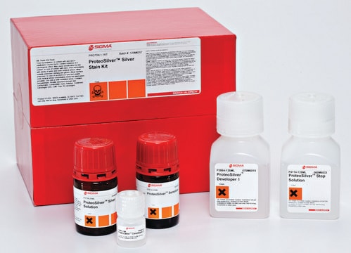
Conveniently packaged, extremely stable, and highly sensitive, ProteoSilver Plus Silver Stain Kit is an ideal product for any protein scientist. Similar to ProteoSilver (PROTSIL1), this kit contains prepared solutions of silver staining reagents along with detailed instructions to achieve optimal results. With a detection limit of 0.1 ng/mm2 of protein (BSA) and an extremely low background, ProteoSilver Plus leads to superior detection of low abundance proteins.
ProteoSilver Plus is also MALDI-MS compatible and does not contain glutaraldehyde, which crosslinks lysine residues, resulting in inaccurate spectra. ProteoSilver Plus Silver Stain Kit contains two additional reagents for destaining an excised protein spot, for those wishing to perform further characterization through tryptic digestion and MALDI-MS analysis.

Application

ProteoSilver Silver Stain Kit has been used for staining of the gels for visualizing proteins. It has also been used for destaining of the protein of interest using destainer provided in the kit for mass spectroscopic analysis.

Features and Benefits

  • Premixed and preweighed solutions reduce time and cost of purchasing and preparing individual components
  • Optimized protocols help establish conditions for best results
  • High sensitivity and low background ensure very low abundance proteins can be detected and resolved from other proteins
  • MALDI Compatible - Protein spots of interest can be further characterized by mass spectrometry
  • Room Temperature Stability allows easy and convenient storage

Legal Information

ProteoSilver is a trademark of Sigma-Aldrich Co. LLC

signalword

Danger

Hazard Classifications

Acute Tox. 2 Inhalation - Acute Tox. 3 Dermal - Acute Tox. 3 Oral - Aquatic Acute 1 - Aquatic Chronic 1 - Carc. 1B - Eye Dam. 1 - Met. Corr. 1 - Muta. 2 - Repr. 1B - Skin Corr. 1A - Skin Sens. 1 - STOT SE 2 - STOT SE 3

target_organs

Eyes,Central nervous system, Respiratory system

Storage Class

6.1A - Combustible acute toxic Cat. 1 and 2 / very toxic hazardous materials

wgk_germany

WGK 3

flash_point_f

149.0 °F - closed cup

flash_point_c

65 °C - closed cup


Choose from one of the most recent versions:

Certificates of Analysis (COA)

Lot/Batch Number

Don't see the Right Version?

If you require a particular version, you can look up a specific certificate by the Lot or Batch number.

Already Own This Product?

Find documentation for the products that you have recently purchased in the Document Library.

Visit the Document Library

Customers Also Viewed

2D map of proteins from human renal stone matrix and evaluation of their effect on oxalate induced renal tubular epithelial cell injury.
Aggarwal KP, et al.
International Brazilian Journal of Urology : Official Journal of the Brazilian Society of Urology, 39, 128-136 (2013)
Effect of biomolecules from human renal matrix of calcium oxalate monohydrate (CaOx) stones on in vitro calcium phosphate crystallization.
Pathak P, et al.
International Brazilian Journal of Urology : Official Journal of the Brazilian Society of Urology, 36, 621-628 (2010)
Kanu Priya Aggarwal et al.
PloS one, 8(7), e69916-e69916 (2013-07-31)
The increasing number of patients suffering from urolithiasis represents one of the major challenges which nephrologists face worldwide today. For enhancing therapeutic outcomes of this disease, the pathogenic basis for the formation of renal stones is the need of hour.
Peeping into human renal calcium oxalate stone matrix: characterization of novel proteins involved in the intricate mechanism of urolithiasis.
Aggarwal KP, et al.
PLoS ONE, 8, e69916-e69916 (2013)
Slovénie Pyndiah et al.
Molecular & cellular proteomics : MCP, 6(2), 193-206 (2006-11-10)
The study of protein interactions constitutes an important domain to understand the physiology and pathogenesis of microorganisms. The two-dimensional blue native/SDS-PAGE was initially reported to analyze membrane protein complexes. In this study, both cytoplasmic and membrane complexes of a bacterium

Articles

To meet the great diversity of protein analysis needs, Sigma offers a wide selection of protein visualization (staining) reagents. EZBlue™ and ProteoSilver™, designed specifically for proteomics, also perform impressively in traditional PAGE formats.

Our team of scientists has experience in all areas of research including Life Science, Material Science, Chemical Synthesis, Chromatography, Analytical and many others.

Contact Technical Service