MABT345
Anti-Prelamin-A Antibody, clone 7G11
clone 7G11, from rat
Synonym(s):
Prelamin-A/C, Prelamin-A, Lamin-A/C
About This Item
Recommended Products
biological source
rat
Quality Level
antibody form
purified antibody
antibody product type
primary antibodies
clone
7G11, monoclonal
species reactivity
mouse
technique(s)
immunofluorescence: suitable
western blot: suitable
isotype
IgG2aκ
NCBI accession no.
UniProt accession no.
shipped in
wet ice
target post-translational modification
unmodified
Gene Information
mouse ... Lmna(16905)
General description
Specificity
Immunogen
Application
Cell Structure
Adhesion (CAMs)
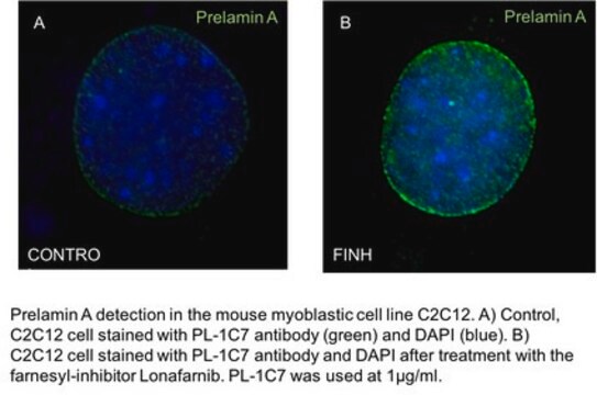
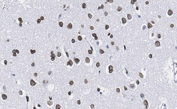
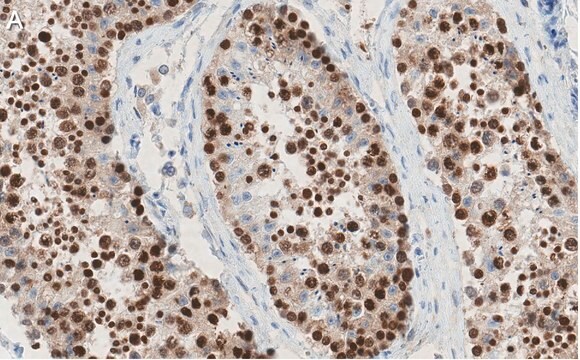
Immunofluorescence Analysis: A representative lot detected upregulated prelamin-A immunoreactivity in keratinocytes, but not in dermal fibroblasts by fluorescent immunohistochemistry using paraformaldehyde-fixed and paraffin-embedded skin sections from keratinocyte-specific Fntb knockout mice, while prelamin-A upregulation was seen in both keratinocytes and dermal fibroblasts from mice with non-tissue/cell type-specific Zmpste24 knockout (Lee, R., et al. (2010). Hum. Mol. Genet.19(8):1603-1617).
Immunofluorescence Analysis: A representative lot stained the nuclear rim of hepatocytes by fluorescent immunohistochemistry using methanol-fixed and Tween-permeabilized frozen liver sections from a transgenic mouse strain that produces the C662S non-farnesylated prelamin-A only (nPLAO/nPLAO) and no lamin-C, while cellular prelamin-A level was undetectable in liver sections from wild-type mice or a mouse strain that produces wild-type prelamin-A only (PLAO/PLAO) and no lamin-C (Davies, B.S., et al. (2010). Hum. Mol. Genet. 19(13):2682-2694).
Western Blotting Analysis: A representative lot detected cellular prelamin-A accumulation upon farnesyltransferase inhibitor (FTI) treatment in murine fibroblasts. Clone 7G11 selectively detects prelamin-A, but not mature lamin-A or lamin-C (Lee, R., et al. (2010). Hum. Mol. Genet.19(8):1603-1617).
Note: Under normal condition, cellular prelamin-A level is below detection. Any newly produced prelamin-A is quickly processed further to yield mature lamin-A. Cellular farnesyltransferase and/or ZMPSTE24 activity must be inhibited (by gene knockout or inhibitor treatment) to allow antibody-based detection of cellular prelamin-A.
Quality
Western Blotting Analysis: 0.5 µg/mL of this antibody detected 2-day FTI-276 (5 µM; Cat. No. 344550) treatment-induced prelamin-A accumulation in murine RAW 264.7 macrophages.
Target description
Physical form
Storage and Stability
Other Notes
Disclaimer
Not finding the right product?
Try our Product Selector Tool.
Storage Class
12 - Non Combustible Liquids
wgk_germany
WGK 1
flash_point_f
Not applicable
flash_point_c
Not applicable
Certificates of Analysis (COA)
Search for Certificates of Analysis (COA) by entering the products Lot/Batch Number. Lot and Batch Numbers can be found on a product’s label following the words ‘Lot’ or ‘Batch’.
Already Own This Product?
Find documentation for the products that you have recently purchased in the Document Library.
Our team of scientists has experience in all areas of research including Life Science, Material Science, Chemical Synthesis, Chromatography, Analytical and many others.
Contact Technical Service