MABS1267
Anti-Nuclear Pore Complex Proteins Antibody, clone 414
clone 414, from mouse
Synonym(s):
Nuclear pore glycoprotein p62, 62 kDa nucleoporin, Nucleoporin Nup62, Nuclear pore complex proteins
About This Item
Recommended Products
biological source
mouse
Quality Level
antibody form
purified immunoglobulin
antibody product type
primary antibodies
clone
414, monoclonal
species reactivity
yeast, mouse, rat, human
technique(s)
electron microscopy: suitable
immunocytochemistry: suitable
immunoprecipitation (IP): suitable
western blot: suitable
isotype
IgG1κ
NCBI accession no.
UniProt accession no.
target post-translational modification
unmodified
Gene Information
human ... NUP62(23636)
General description
Specificity
Immunogen
Application
Immunocytochemistry Analysis: A representative lot immunostained yeast nuclear envelope with a punctate and patchy pattern (Aris, J.P., and Blobel, G. (1989). J. Cell Biol. 108(6):2059-2067).
Immunocytochemistry Analysis: Representative lots detected a punctate staining pattern of Nup62 at the nuclear rim by fluorescent immunocytochemistry using 2% formaldehyde-fixed, methanol-permeabilized Buffalo rat liver (BRL) cells (Davis, L.I., and Blobel, G. (1987). Proc. Natl. Acad. Sci. U. S. A. 84(21):7552-755; Davis, L.I., and Blobel, G. (1986). Cell. 45(5):699-709).
Electron Microscopy: A representative lot immunostained yeast nuclear envelope using 3% paraformaldehyde/0.2% glutaraldehyde-fixed yeast nuclei LR White sections (Aris, J.P., and Blobel, G. (1989). J. Cell Biol. 108(6):2059-2067).
Electron Microscopy: A representative lot immunostained the pore complexes in thin sections of isolated rat liver nuclei extracted with 2% Triton X-100 and fixed with 0.05% glutaraldehyde (Davis, L.I., and Blobel, G. (1986). Cell. 45(5):699-709).
Immunoprecipitation Analysis: A representative lot immunoprecipited a ~100 kDa (p110) and a ~95 kDa (p95) protein species from yeast nuclear extract. An additional ~55 kDa protein was immunoprecipitated by clone 414 using yeast cytosolic fraction or whole cell lysate (Aris, J.P., and Blobel, G. (1989). J. Cell Biol. 108(6):2059-2067).
Immunoprecipitation Analysis: A representative lot immunoprecipitated three major GlcNAcylated prtoein species of 62, 175, and 270 kDa from SDS-solubilized pore complex-lamina extract of Buffalo rat liver (BRL) cell nuclei preparation that had been labeled with UDP-[3H]Gal by galactosyltransferase (Davis, L.I., and Blobel, G. (1987). Proc. Natl. Acad. Sci. U. S. A. 84(21):7552-755).
Immunoprecipitation Analysis: A representative lot immunoprecipitated GlcNAcylated 62 kDa protein (p62) from the SDS-solubilized pore complex-lamina extract, as well as a less glycosylated p61 cytoplasmic form from the postmitochondrial supernatant of Buffalo rat liver (BRL) cells (7552-755; Davis, L.I., and Blobel, G. (1986). Cell. 45(5):699-709).
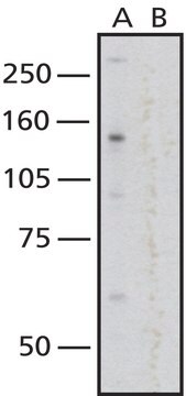
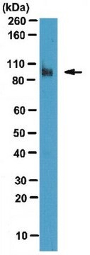
Western Blotting Analysis: A representative lot detected a ~100 kDa (p110) and a ~95 kDa (p95) immunoreactive bands in yeast nuclear extract (Aris, J.P., and Blobel, G. (1989). J. Cell Biol. 108(6):2059-2067).
Western Blotting Analysis: A representative lot detected a ~62 kDa (p62) and a ~200 kDa target bands associated with nuclear pore complex-lamina of rat liver nuclei preparation even following sequential nucleaases, Triton X-100, and 140 mM NaCl treatments (Davis, L.I., and Blobel, G. (1986). Cell. 45(5):699-709).
Signaling
Chromatin Biology
Quality
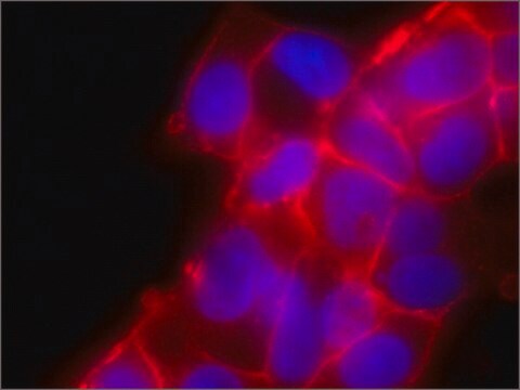
Immunocytochemistry Analysis: A 1:200 dilution of this antibody immunostained HeLa cell nuclear rim.
Target description
Physical form
Storage and Stability
Other Notes
Disclaimer
Not finding the right product?
Try our Product Selector Tool.
Storage Class
12 - Non Combustible Liquids
wgk_germany
WGK 1
flash_point_f
Not applicable
flash_point_c
Not applicable
Certificates of Analysis (COA)
Search for Certificates of Analysis (COA) by entering the products Lot/Batch Number. Lot and Batch Numbers can be found on a product’s label following the words ‘Lot’ or ‘Batch’.
Already Own This Product?
Find documentation for the products that you have recently purchased in the Document Library.
Our team of scientists has experience in all areas of research including Life Science, Material Science, Chemical Synthesis, Chromatography, Analytical and many others.
Contact Technical Service