Skip to Content
MilliporeSigma
All Photos(2)

Key Documents

04-886

Sigma-Aldrich

Anti-phospho-STAT5A/B (Tyr694/699) Antibody, clone A11W, rabbit monoclonal

culture supernatant, clone A11W, from rabbit

Synonym(s):

signal transducer and activator of transcription 5A, signal transducer and activator of transcription 5B, transcription factor STAT5B

Sign Into View Organizational & Contract Pricing


About This Item

UNSPSC Code:
12352203
eCl@ss:
32160702
NACRES:
NA.41
clone:
A11W, monoclonal
application:
WB
species reactivity:
rat, chimpanzee, human, mouse, bovine
technique(s):
western blot: suitable
citations:
6

biological source

rabbit

Quality Level

antibody form

culture supernatant

antibody product type

primary antibodies

clone

A11W, monoclonal

species reactivity

rat, chimpanzee, human, mouse, bovine

technique(s)

western blot: suitable

isotype

IgG

suitability

not suitable for immunoprecipitation

NCBI accession no.

UniProt accession no.

shipped in

dry ice

target post-translational modification

phosphorylation (pTyr694/pTyr699)

Gene Information

bovine ... Stat5A(282375)

General description

The STAT proteins (signal transducer and activator of transcription) comprise a family of transcription factors. STATs are activated through tyrosine phosphorylation, mainly by JAK kinases, which results in their dimerization, nuclear translocation and regulation of gene expression. STAT5 activity is induced by numerous cytokines and growth factors. While STAT5A and STAT5B share 96% amino acid homology, the two isoforms have distinct physiological functions. Phosphorylation of tyrosine 694/699 is a key marker of STAT5A/B activation.

Specificity

Phospho-STAT5A/B (Tyr694/699).

Immunogen

Epitope: pTyr694/699
KLH-conjugated, synthetic peptide containing the sequence DGpYVK in which pY corresponds to phospho-tyrosine at residue 694 of human STAT5A or residue 699 of STAT5B.

Application

Detect phospho-STAT5A/B (Tyr694/699) using this Anti-phospho-STAT5A/B (Tyr694/699) Antibody, clone A11W validated for use in WB.
Research Category
Signaling

Apoptosis & Cancer
Research Sub Category
Transcription Factors

Quality

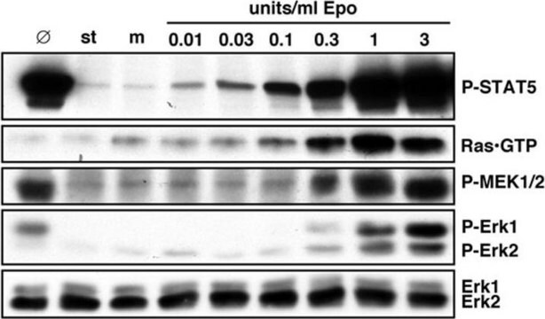
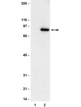
Evaluated by western blot on RIPA lysates from 3T3/NIH cells untreated and treated with PDGF.

Western Blot Analysis:
A 1:1,000-1:2,000 dilution of this antibody detected phospho-STAT5A/B (Tyr694/699) in lysates from 3T3/NIH cells treated with PDGF.

Target description

~95 kDa

Linkage

Replaces: 05-886

Physical form

Cultured supernatant containing 0.05% sodium azide.

Storage and Stability

Stable for 1 year at -20ºC from date of receipt.
Handling Recommendations: Upon receipt, and prior to removing the cap, centrifuge the vial and gently mix the solution. Aliquot into microcentrifuge tubes and store at -20°C. Avoid repeated freeze/thaw cycles, which may damage IgG and affect product performance.

Analysis Note

Control
PDGF treated 3T3/NIH cells

Disclaimer

Unless otherwise stated in our catalog or other company documentation accompanying the product(s), our products are intended for research use only and are not to be used for any other purpose, which includes but is not limited to, unauthorized commercial uses, in vitro diagnostic uses, ex vivo or in vivo therapeutic uses or any type of consumption or application to humans or animals.

Not finding the right product?  

Try our Product Selector Tool.

Storage Class

12 - Non Combustible Liquids

wgk_germany

WGK 1

flash_point_f

Not applicable

flash_point_c

Not applicable


Certificates of Analysis (COA)

Search for Certificates of Analysis (COA) by entering the products Lot/Batch Number. Lot and Batch Numbers can be found on a product’s label following the words ‘Lot’ or ‘Batch’.

Already Own This Product?

Find documentation for the products that you have recently purchased in the Document Library.

Visit the Document Library

Altered interleukin-12 responsiveness in Th1 and Th2 cells is associated with the differential activation of STAT5 and STAT1.
Gollob, J A, et al.
Blood, 91, 1341-1354 (1998)
Pey-Jium Chang et al.
Oncotarget, 8(46), 80595-80611 (2017-11-09)
Patients with diabetes are generally prone to pathogen infection and tumor progression. Here, we investigated the potential association between diabetes and Kaposi's sarcoma (KS), a tumor linked to infection with Kaposi's sarcoma-associated herpesvirus (KSHV). By using Taiwan's National Health Insurance
John F Woolley et al.
PloS one, 7(7), e34050-e34050 (2012-07-19)
The internal tandem duplication (ITD) of the juxtamembrane region of the FLT3 receptor has been associated with increased reactive oxygen species (ROS) generation in acute myeloid leukemia (AML). How this elevated level of ROS contributes to the leukemic phenotype, however

Our team of scientists has experience in all areas of research including Life Science, Material Science, Chemical Synthesis, Chromatography, Analytical and many others.

Contact Technical Service