Przejdź do zawartości
Merck

SAB4200742

Sigma-Aldrich

Anti-c-Myc-Peroxidase antibody, Mouse monoclonal

clone 9E10, purified from hybridoma cell culture

Synonim(y):

Anti-MRTL, Anti-Myc, Anti-bHLHe39, Anti-v-myc avian myelocytomatosis viral oncogene homolog

Zaloguj sięWyświetlanie cen organizacyjnych i kontraktowych


About This Item

Kod UNSPSC:
12352203
NACRES:
NA.56

pochodzenie biologiczne

mouse

Poziom jakości

białko sprzężone

peroxidase conjugate

forma przeciwciała

purified from hybridoma cell culture

rodzaj przeciwciała

primary antibodies

klon

9E10, monoclonal

Formularz

lyophilized powder

reaktywność gatunkowa

human

opakowanie

vial of 100 μL

stężenie

~2 mg/mL

metody

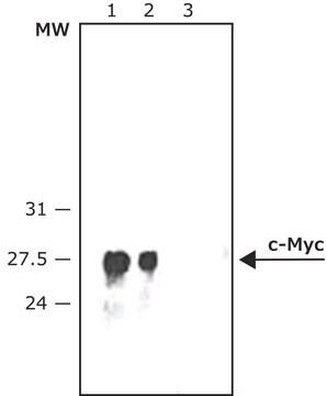
immunoblotting: 1:250-1:500 using lysate of HEK-293T cells over expressing c-Myc fusion protein
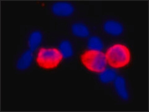
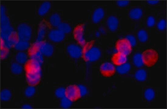
immunohistochemistry: suitable

izotyp

IgG1

numer dostępu UniProt

Warunki transportu

dry ice

temp. przechowywania

−20°C

docelowa modyfikacja potranslacyjna

unmodified

informacje o genach

human ... MYC(4609)

Opis ogólny

Monoclonal Anti-c-Myc (mouse IgG1 isotype) is derived from the 9E10 hybridoma, produced by the fusion of mouse myeloma cells and splenocytes from a BALB/c mouse immunized with synthetic peptide of the human p62c-myc protein, conjugated to KLH. c-Myc is a proto-oncogene localized on long arm of human chromosome 8. The encoded protein contains an N-terminal transactivation domain. The human c-Myc proto-oncogene is the cellular homolog of the avian v-Myc gene found in several leukemogenic retroviruses.

Specyficzność

Monoclonal Anti-Calponin specifically recognizes smooth muscle Calponin.

Immunogen

Synthetic peptide of the human p62c-myc protein.

Zastosowanie

Anti-c-Myc-Peroxidase antibody, Mouse monoclonal may be used in immunoblotting and immunohistology.

Działania biochem./fizjol.

c-Myc is a transcription factor involved in regulation of cell growth, proliferation, differentiation and apoptosis. It is implicated in DNA replication, RNA splicing or transcription. Increased expression of the gene is associated with the development of various types of human cancers.

Postać fizyczna

Supplied as lyophilized powder. After reconstitution with 0.1 mL of distilled water to a final antibody concentration of ∼ 2 mg/mL, the solution contains 1% BSA, 2.5% trehalose, 0.05% MIT in 0.01 M sodium phosphate buffered saline

Przechowywanie i stabilność

Store the lyophilized product at 2–8 °C. For extended storage after reconstitution, keep at –20 °C in working aliquots. Avoid repeated freeze-thaw cycles. For continuous use after reconstitution, keep at 2–8 °C for up to 1 month. Solutions at working dilution should be discarded if not used within 12 hours.

Oświadczenie o zrzeczeniu się odpowiedzialności

Unless otherwise stated in our catalog, our products are intended for research use only and are not to be used for any other purpose, which includes but is not limited to, unauthorized commercial uses, in vitro diagnostic uses, ex vivo or in vivo therapeutic uses or any type of consumption or application to humans or animals.
Ta strona może zawierać tekst przetłumaczony maszynowo.

Nie możesz znaleźć właściwego produktu?  

Wypróbuj nasz Narzędzie selektora produktów.

Piktogramy

Exclamation mark

Hasło ostrzegawcze

Warning

Zwroty wskazujące rodzaj zagrożenia

Zwroty wskazujące środki ostrożności

Klasyfikacja zagrożeń

Skin Sens. 1

Kod klasy składowania

13 - Non Combustible Solids

Klasa zagrożenia wodnego (WGK)

WGK 2

Temperatura zapłonu (°F)

Not applicable

Temperatura zapłonu (°C)

Not applicable


Wybierz jedną z najnowszych wersji:

Certyfikaty analizy (CoA)

Lot/Batch Number

Nie widzisz odpowiedniej wersji?

Jeśli potrzebujesz konkretnej wersji, możesz wyszukać konkretny certyfikat według numeru partii lub serii.

Masz już ten produkt?

Dokumenty związane z niedawno zakupionymi produktami zostały zamieszczone w Bibliotece dokumentów.

Odwiedź Bibliotekę dokumentów

K Alitalo et al.
Nature, 306(5940), 274-277 (1983-11-17)
The myelocytomatosis viruses are a family of replication-defective avian retroviruses that cause a variety of tumours in chickens and transform both fibroblasts and macrophages in culture through the activity of their oncogene v-myc. A closely related gene (c-myc) is found
S Pelengaris et al.
Current opinion in genetics & development, 10(1), 100-105 (2000-02-19)
The protein products of many dominant oncogenes are capable of inducing both cell proliferation and apoptosis. Recent experiments employing transgenic mice that express an ectopically regulatable myc gene or protein have begun to elucidate the role of the balance between
Q Hu et al.
Science (New York, N.Y.), 268(5207), 100-102 (1995-04-07)
Phosphatidylinositol (Pl)-3 kinase is one of many enzymes stimulated by growth factors. A constitutively activated mutant, p110, that functions independently of growth factor stimulation was constructed to determine the specific responses regulated by Pl-3 kinase. The p110 protein exhibited high
Ann-Marie Baker et al.
Histopathology, 69(2), 222-229 (2016-01-31)
Recent attempts to study MYC distribution in human samples have been confounded by a lack of agreement in immunohistochemical staining between antibodies targeting the N-terminus and those targeting the C-terminus of the MYC protein. The aim of this study was
D Robertson et al.
The journal of histochemistry and cytochemistry : official journal of the Histochemistry Society, 43(5), 471-480 (1995-05-01)
To determine the ultrastructural distribution of H-ras, the rho proteins rho-A, rho-B, rho-C, and the rac1 protein (members of the ras GTP-binding protein family), we used cDNA expression plasmids in which a short sequence coding for the epitope recognized by

Nasz zespół naukowców ma doświadczenie we wszystkich obszarach badań, w tym w naukach przyrodniczych, materiałoznawstwie, syntezie chemicznej, chromatografii, analityce i wielu innych dziedzinach.

Skontaktuj się z zespołem ds. pomocy technicznej