Przejdź do zawartości
Merck
Wszystkie zdjęcia(1)

Kluczowe dokumenty

369A-1

Sigma-Aldrich

Phosphohistone H3 (PHH3) Rabbit Polyclonal Antibody

Zaloguj sięWyświetlanie cen organizacyjnych i kontraktowych


About This Item

Kod UNSPSC:
12352203
NACRES:
NA.41

pochodzenie biologiczne

rabbit

Poziom jakości

100
500

białko sprzężone

unconjugated

forma przeciwciała

Ig fraction of antiserum

rodzaj przeciwciała

primary antibodies

klon

polyclonal

opis

For In Vitro Diagnostic Use in Select Regions (See Chart)

Formularz

buffered aqueous solution

reaktywność gatunkowa

human

opakowanie

vial of 0.1 mL concentrate (369A-14)
vial of 0.5 mL concentrate (369A-15)
bottle of 1.0 mL predilute (369A-17)
vial of 1.0 mL concentrate (369A-16)
bottle of 7.0 mL predilute (369A-18)

producent / nazwa handlowa

Cell Marque

metody

immunohistochemistry (formalin-fixed, paraffin-embedded sections): 1:100-1:500

kontrola

tonsil

Warunki transportu

wet ice

temp. przechowywania

2-8°C

wizualizacja

nuclear

informacje o genach

human ... H3C1(8350)

Opis ogólny

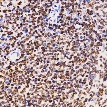
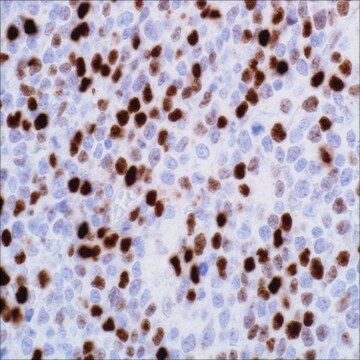
Phosphohistone H3 (PHH3) is a core histone protein, which together with other histones, forms the major protein constituents of the chromatin in eukaryotic cells. In mammalian cells, phosphohistone H3 is negligible during interphase but reaches a maximum for chromatin condensation during mitosis. Immunohistochemical studies showed anti-PHH3 specifically detected the core protein histone H3 only when phosphorylated at serine 10 or serine 28. Studies have also revealed no phosphorylation on the histone H3 during apoptosis. Therefore, PHH3 can serve as a mitotic marker to separate mitotic figures from apoptotic bodies and karyorrhectic debris, which may be a very useful tool in the diagnosis of tumor grades, especially in CNS, skin, gyn., soft tissue, and GIST.

Jakość


IVD

IVD

IVD

RUO

Powiązanie

Phosphohistone H3 (PHH3) Positive Control Slides, Product No. 369S, are available for immunohistochemistry (formalin-fixed, paraffin-embedded sections).

Postać fizyczna

Solution in Tris Buffer, pH 7.3-7.7, with 1% BSA and <0.1% Sodium Azide

Uwaga dotycząca przygotowania

Download the IFU specific to your product lot and formatNote: This requires a keycode which can be found on your packaging or product label.

Inne uwagi

For Technical Service please contact: 800-665-7284 or email: service@cellmarque.com

Informacje prawne

Cell Marque is a trademark of Merck KGaA, Darmstadt, Germany
Ta strona może zawierać tekst przetłumaczony maszynowo.

Nie możesz znaleźć właściwego produktu?  

Wypróbuj nasz Narzędzie selektora produktów.


Wybierz jedną z najnowszych wersji:

Certyfikaty analizy (CoA)

Lot/Batch Number

Nie widzisz odpowiedniej wersji?

Jeśli potrzebujesz konkretnej wersji, możesz wyszukać konkretny certyfikat według numeru partii lub serii.

Masz już ten produkt?

Dokumenty związane z niedawno zakupionymi produktami zostały zamieszczone w Bibliotece dokumentów.

Odwiedź Bibliotekę dokumentów

M J Hendzel et al.
The Journal of biological chemistry, 273(38), 24470-24478 (1998-09-12)
Apoptosis plays an important role in the survival of an organism, and substantial work has been done to understand the signaling pathways that regulate this process. Characteristic changes in chromatin organization accompany apoptosis and are routinely used as markers for
Histone phosphorylation and chromatin structure during mitosis in Chinese hamster cells.
L R Gurley et al.
European journal of biochemistry, 84(1), 1-15 (1978-03-01)
Michel R Nasr et al.
The American Journal of dermatopathology, 30(2), 117-122 (2008-03-25)
Differentiating malignant melanoma from benign melanocytic lesions can be challenging. We undertook this study to evaluate the use of the immunohistochemical mitosis marker phospho-Histone H3 (pHH3) and the proliferation markers Ki-67 and survivin in separating malignant melanoma from benign nevi.
Howard Colman et al.
The American journal of surgical pathology, 30(5), 657-664 (2006-05-16)
Distinguishing between grade II and grade III diffuse astrocytomas is important both for prognosis and for treatment decision-making. However, current methods for distinguishing between grades based on proliferative potential are suboptimal, making identification of clear cutoffs difficult. In this study
Yoo-Jin Kim et al.
American journal of clinical pathology, 128(1), 118-125 (2007-06-21)
Mitotic activity is one of the most reliable prognostic factors in meningiomas. The identification of mitotic figures (MFs) and the areas of highest mitotic activity in H&E-stained slides is a tedious and subjective task. Therefore, we compared the results from

Nasz zespół naukowców ma doświadczenie we wszystkich obszarach badań, w tym w naukach przyrodniczych, materiałoznawstwie, syntezie chemicznej, chromatografii, analityce i wielu innych dziedzinach.

Skontaktuj się z zespołem ds. pomocy technicznej