Przejdź do zawartości
Merck
Wszystkie zdjęcia(3)

Kluczowe dokumenty

MABN495

Sigma-Aldrich

Anti-Aldh1L1 Antibody, clone N103/39

clone N103/39, from mouse

Synonim(y):

Cytosolic 10-formyltetrahydrofolate dehydrogenase, 10-FTHFDH, FDH, Aldehyde dehydrogenase family 1 member L1

FBP-CI

Zaloguj sięWyświetlanie cen organizacyjnych i kontraktowych


About This Item

Kod UNSPSC:
12352203
eCl@ss:
32160702
NACRES:
NA.41

pochodzenie biologiczne

mouse

Poziom jakości

forma przeciwciała

purified immunoglobulin

rodzaj przeciwciała

primary antibodies

klon

N103/39, monoclonal

reaktywność gatunkowa

human, rat, mouse

metody

immunofluorescence: suitable
immunohistochemistry: suitable
western blot: suitable

izotyp

IgG1κ

numer dostępu NCBI

numer dostępu UniProt

Warunki transportu

wet ice

docelowa modyfikacja potranslacyjna

unmodified

informacje o genach

human ... ALDH1L1(10840)

Opis ogólny

Aldh1L1, also known as 10-formyltetrahydrofolate dehydrogenase (10-FTHFDH), is a multidomain protein that is found in liver cytosol and also serves as a CNS astrocyte marker. Aldh1L1 is comprised of two functionally unrelated domains, an aldehyde dehydrogenase-homologous domain and a folate-binding hydrolase domain. These two domains are thought to be connected by a 100-residue linker. It is known to induce phosphorylation of p53 at Ser6, which is a critical step in stimulating apoptosis.

Specyficzność

This Aldh1L1 monoclonal antibody does not exhibit cross-reactivity toward Aldh1L2.

Immunogen

Recombinant full-length rat Aldh1L1.

Zastosowanie

Anti-Aldh1L1, clone N103/39 is an antibody targeting the Aldh1L1 protein, validated for use in Immunofluorescence, Immunohistochemistry, and Western Blotting.
Research Category
Neuroscience
Research Sub Category
Sensory & PNS
Western Blot Analysis: 0.5 µg/mL from a representative lot detected Aldh1L1 in 10 µg of human brain tissue lysate.

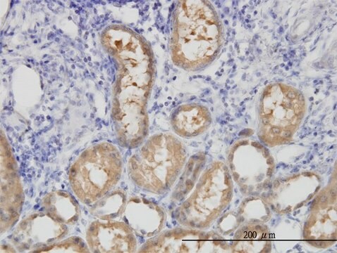
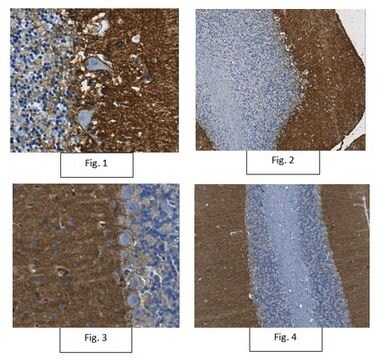
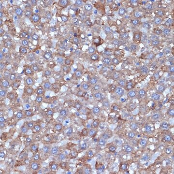
Immunohistochemistry Analysis: A 1:2,000 dilution from a representative lot detected Aldh1L1 rat cerebral cortex tissue and human pons/midbrain tissue.

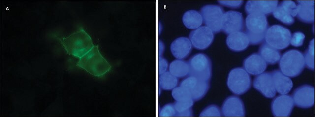
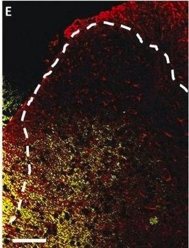
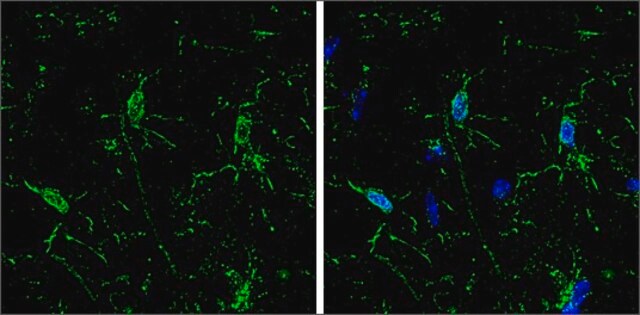
Immunofluorescence Analysis: A representative lot detected Aldh1L1 in rat cortex and cerebellum tissue.

Jakość

Evaluated by Western Blot in mouse brain tissue lysate.

Western Blot Analysis: 0.5 µg/mL of this antibody detected Aldh1L1 in 10 µg of mouse brain tissue lysate.

Opis wartości docelowych

~99 kDa observed

Postać fizyczna

Format: Purified
Protein G Purified
Purified mouse IgG1κ in buffer containing 0.1 M Tris-Glycine (pH 7.4), 150 mM NaCl with 0.05% sodium azide.

Przechowywanie i stabilność

Stable for 1 year at 2-8°C from date of receipt.

Komentarz do analizy

Control
Mouse brain tissue lysate

Inne uwagi

Concentration: Please refer to the Certificate of Analysis for the lot-specific concentration.

Oświadczenie o zrzeczeniu się odpowiedzialności

Unless otherwise stated in our catalog or other company documentation accompanying the product(s), our products are intended for research use only and are not to be used for any other purpose, which includes but is not limited to, unauthorized commercial uses, in vitro diagnostic uses, ex vivo or in vivo therapeutic uses or any type of consumption or application to humans or animals.
Ta strona może zawierać tekst przetłumaczony maszynowo.

Nie możesz znaleźć właściwego produktu?  

Wypróbuj nasz Narzędzie selektora produktów.

Kod klasy składowania

12 - Non Combustible Liquids

Klasa zagrożenia wodnego (WGK)

WGK 1

Temperatura zapłonu (°F)

Not applicable

Temperatura zapłonu (°C)

Not applicable


Certyfikaty analizy (CoA)

Poszukaj Certyfikaty analizy (CoA), wpisując numer partii/serii produktów. Numery serii i partii można znaleźć na etykiecie produktu po słowach „seria” lub „partia”.

Masz już ten produkt?

Dokumenty związane z niedawno zakupionymi produktami zostały zamieszczone w Bibliotece dokumentów.

Odwiedź Bibliotekę dokumentów

Maria de Los Angeles Castillo-Rodriguez et al.
Histochemistry and cell biology, 158(1), 15-38 (2022-04-06)
Myelin loss with consecutive axon degeneration and impaired remyelination are the underlying causes of progressive disease in patients with multiple sclerosis. Astrocytes are suggested to play a major role in these processes. The unmasking of distinct astrocyte identities in health
New ?- and ?-synuclein immunopathological lesions in human brain.
Surgucheva, I; Newell, KL; Burns, J; Surguchov, A
Acta Neuropathologica Communications null
Gianluca L Russo et al.
Cell stem cell, 28(3), 524-534 (2020-11-18)
Astrocyte-to-neuron conversion is a promising avenue for neuronal replacement therapy. Neurons are particularly dependent on mitochondrial function, but how well mitochondria adapt to the new fate is unknown. Here, we determined the comprehensive mitochondrial proteome of cortical astrocytes and neurons
Lindsay T Michalovicz et al.
Journal of neurochemistry, 150(4), 420-440 (2019-06-22)
Neurotoxicology is hampered by the inability to predict regional and cellular targets of toxicant-induced damage. Evaluating astrogliosis overcomes this problem because reactive astrocytes highlight the location of toxicant-induced damage. While enhanced expression of glial fibrillary acidic protein is a hallmark
Krishna C Vadodaria et al.
Stem cell reports, 16(4), 825-835 (2021-03-06)
Bipolar disorder (BD) is characterized by cyclical mood shifts. Studies indicate that BD patients have a peripheral pro-inflammatory state and alterations in glial populations in the brain. We utilized an in vitro model to study inflammation-related phenotypes of astrocytes derived from

Nasz zespół naukowców ma doświadczenie we wszystkich obszarach badań, w tym w naukach przyrodniczych, materiałoznawstwie, syntezie chemicznej, chromatografii, analityce i wielu innych dziedzinach.

Skontaktuj się z zespołem ds. pomocy technicznej