MABF2804
Anti-Reticular fibroblasts Antibody, clone ER-TR7
Synonim(y):
Fibroblast marker
About This Item
IHC (p)
immunohistochemistry (formalin-fixed, paraffin-embedded sections): suitable
Polecane produkty
pochodzenie biologiczne
rat
Poziom jakości
białko sprzężone
unconjugated
forma przeciwciała
purified antibody
rodzaj przeciwciała
primary antibodies
klon
ER-TR7, monoclonal
oczyszczone przez
using protein G
reaktywność gatunkowa
mouse, human
opakowanie
antibody small pack of 100 μg
metody
immunofluorescence: suitable
immunohistochemistry (formalin-fixed, paraffin-embedded sections): suitable
izotyp
IgG2a
sekwencja epitopowa
Unknown
numer dostępu UniProt
Warunki transportu
2-8°C
docelowa modyfikacja potranslacyjna
unmodified
Opis ogólny
Specyficzność
Immunogen
Zastosowanie
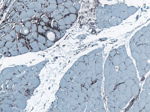
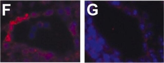
Evaluated by Immunohistochemistry in Mouse thymus tissue sections.
Immunohistochemistry Applications: A 1:50 dilution of this antibody detected reticular fibroblasts in frozen section of Mouse thymus tissue.
Tested applications
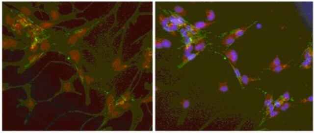
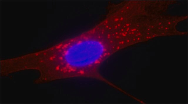
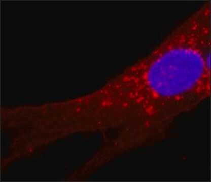
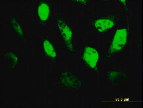
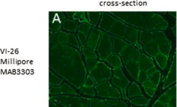
Immunofluorescence Analysis: A representative lot detected Reticular fibroblasts clone ER-TR7 in Immunofluorescence applications (Van Vliet, E., et al. (1984). Eur J Immunol. 14(6):524-9; Nolte, M.A., et al. (2003). J Exp Med. 198(3):505-12; Sixt, M., et al. (2005). Immunity. 22(1):19-29; Link, A., et al. (2007). Nat Immunol. 8(11):1255-65; Odaka, C., et al. (2009). J Histochem Cytochem. 57(4):373-82; Schiavinato, A., et al. (2021). Eur J Immunol. 51(9):2345-2347).
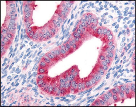
Immunohistochemistry Applications: A representative lot detected Reticular fibroblasts clone ER-TR7 in Immunohistochemistry applications (Van Vliet, E., et al. (1984). Eur J Immunol. 14(6):524-9; Nolte, M.A., et al. (2003). J Exp Med. 198(3):505-12; Sixt, M., et al. (2005). Immunity. 22(1):19-29; Link, A., et al. (2007). Nat Immunol.;8(11):1255-65; Odaka, C., et al. (2009). J Histochem Cytochem. 57(4):373-82; Schiavinato, A., et al. (2021). Eur J Immunol. 51(9):2345-2347).
Note: Actual optimal working dilutions must be determined by end user as specimens, and experimental conditions may vary with the end user
Postać fizyczna
Przechowywanie i stabilność
Inne uwagi
Oświadczenie o zrzeczeniu się odpowiedzialności
Nie możesz znaleźć właściwego produktu?
Wypróbuj nasz Narzędzie selektora produktów.
polecane
Kod klasy składowania
12 - Non Combustible Liquids
Klasa zagrożenia wodnego (WGK)
WGK 1
Temperatura zapłonu (°F)
Not applicable
Temperatura zapłonu (°C)
Not applicable
Certyfikaty analizy (CoA)
Poszukaj Certyfikaty analizy (CoA), wpisując numer partii/serii produktów. Numery serii i partii można znaleźć na etykiecie produktu po słowach „seria” lub „partia”.
Masz już ten produkt?
Dokumenty związane z niedawno zakupionymi produktami zostały zamieszczone w Bibliotece dokumentów.
Nasz zespół naukowców ma doświadczenie we wszystkich obszarach badań, w tym w naukach przyrodniczych, materiałoznawstwie, syntezie chemicznej, chromatografii, analityce i wielu innych dziedzinach.
Skontaktuj się z zespołem ds. pomocy technicznej