Przejdź do zawartości
Merck
Wszystkie zdjęcia(1)

Kluczowe dokumenty

MABF2778

Sigma-Aldrich

Anti-IRF-3 Antibody, clone AR-1

Zaloguj sięWyświetlanie cen organizacyjnych i kontraktowych


About This Item

Kod UNSPSC:
12352203
NACRES:
NA.41

pochodzenie biologiczne

mouse

Poziom jakości

białko sprzężone

unconjugated

forma przeciwciała

purified antibody

rodzaj przeciwciała

primary antibodies

klon

AR-1, monoclonal

masa cząsteczkowa

calculated mol wt 47.22 kDa
observed mol wt ~51 kDa

reaktywność gatunkowa

rhesus macaque, human

opakowanie

antibody small pack of 100 μL

metody

ELISA: suitable
flow cytometry: suitable
immunoprecipitation (IP): suitable
inhibition assay: suitable
western blot: suitable

izotyp

IgG1

numer dostępu UniProt

Warunki transportu

dry ice

temp. przechowywania

2-8°C

docelowa modyfikacja potranslacyjna

unmodified

Opis ogólny

Interferon regulatory factor 3 (UniProt: Q14653; also known as IRF-3) is encoded by the IRF3 gene (Gene ID: 3661) in human. IRF-3 is a transcription factor that is constitutively expressed in a variety of tissues and plays a role in intracellular immune response against DNA and RNA viruses. It regulates the transcription of type I interferon genes (IFN-a and IFN-b) and interferon-stimulated genes by binding to an interferon-stimulated response element in their promoters. IRF-3 contains a DNA-binding domain (aa 5-111), nuclear export signal (aa 139-149), IRF-interacting domain, and a C-terminal serine-rich region that contains several phosphorylation sites. Although some of these serines are shown to be phosphorylated in the resting state, further phosphorylation of specific residues is required for its activation and nuclear translocation. In uninfected cells, IRF-3 is present in the cytoplasm, however, following infection, it undergoes phosphorylation on serine 385, serine 386, threonine 390, and serine 396. These phosphorylation events induce a conformational change that leads to its dimerization and nuclear localization where it associates with CREB binding protein to form dsRNA activated factor 1 that subsequently leads to the activation of type I interferons and ISG genes. Phosphorylation and subsequent activation of IRF-3 shown to be inhibited by vaccinia virus protein E3. Mutations in IRF3 gene have been linked to infection-induced, Herpes-specific acute encephalopathy that is characterized by hemorrhagic necrosis of parts of the temporal and frontal lobes. Clone AR-1 recognizes an epitope within the DNA-binding domain of IRF-3. It can detect resting IRF-3 as well as the activated form. (Ref.: Rustagi, A., et al. (2013). Methods. 59(2); 225-232; Honda, K., et al. (2006). Immunity. 25(3); 349-360).

Specyficzność

Clone AR1 is a mouse monoclonal antibody that detects Interferon regulatory factor 3 (IRF-3). It targets an epitope within the DNA-binding domain from the N-terminal half.

Immunogen

His-tagged full-length human recombinant human Interferon regulatory factor 3 (IRF-3).

Zastosowanie

Quality Control Testing

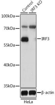
Evaluated by Western Blotting in A549 cell lysate.

Western Blotting Analysis: A 1:500 dilution of this antibody detected IRF-3 in A549 cell lysate.

Tested Applications

ELISA Analysis: A representative lot detected IRF-3 in ELISA applications (Rustagi, A., et. al. (2013). Methods. 59(2):225-32).

Flow Cytometry Analysis: A representative lot detected IRF-3 in Flow Cytometry applications (Rustagi, A., et. al. (2013). Methods. 59(2):225-32).

Western Blotting Analysis: A representative lot detected IRF-3 in Western Blotting applications (Rustagi, A., et. al. (2013). Methods. 59(2):225-32).

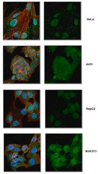
Immunocytochemistry Analysis: A representative lot detected IRF-3 in Immunocytochemistry applications (Rustagi, A., et. al. (2013). Methods. 59(2):225-32).

Note: Actual optimal working dilutions must be determined by end user as specimens, and experimental conditions may vary with the end user
Anti-IRF-3, clone AR-1, Cat. No. MABF2778, is a mouse monoclonal antibody that detects Interferon regulatory factor 3 and is tested for use in ELISA, Flow Cytometry, Immunocytochemistry, and Western Blotting.

Postać fizyczna

Purified mouse monoclonal antibody IgG1 in buffer containing 0.1 M Tris-Glycine (pH 7.4), 150 mM NaCl with 0.05% sodium azide.

Przechowywanie i stabilność

Recommend storage at +2°C to +8°C. For long term storage antibodies can be kept at -20°C. Avoid repeated freeze-thaws.

Inne uwagi

Concentration: Please refer to the Certificate of Analysis for the lot-specific concentration.

Oświadczenie o zrzeczeniu się odpowiedzialności

Unless otherwise stated in our catalog or other company documentation accompanying the product(s), our products are intended for research use only and are not to be used for any other purpose, which includes but is not limited to, unauthorized commercial uses, in vitro diagnostic uses, ex vivo or in vivo therapeutic uses or any type of consumption or application to humans or animals.
Ta strona może zawierać tekst przetłumaczony maszynowo.

Nie możesz znaleźć właściwego produktu?  

Wypróbuj nasz Narzędzie selektora produktów.

Kod klasy składowania

12 - Non Combustible Liquids

Klasa zagrożenia wodnego (WGK)

WGK 1

Temperatura zapłonu (°F)

Not applicable

Temperatura zapłonu (°C)

Not applicable


Certyfikaty analizy (CoA)

Poszukaj Certyfikaty analizy (CoA), wpisując numer partii/serii produktów. Numery serii i partii można znaleźć na etykiecie produktu po słowach „seria” lub „partia”.

Masz już ten produkt?

Dokumenty związane z niedawno zakupionymi produktami zostały zamieszczone w Bibliotece dokumentów.

Odwiedź Bibliotekę dokumentów

Nasz zespół naukowców ma doświadczenie we wszystkich obszarach badań, w tym w naukach przyrodniczych, materiałoznawstwie, syntezie chemicznej, chromatografii, analityce i wielu innych dziedzinach.

Skontaktuj się z zespołem ds. pomocy technicznej