
ABN991
Anti-phospho GLUT-1 Antibody (Ser226)
from rabbit, purified by affinity chromatography
Synonim(y):
Solute carrier family 2, facilitated glucose transporter member 1, Glucose transporter type 1, erythrocyte/brain, GLUT-1, HepG2 glucose transporter, Human T-cell leukemia virus I and II receptor, Receptor for HTLV-1 and HTLV-2, phospho GLUT-1 (Ser226)
About This Item
Polecane produkty


pochodzenie biologiczne
rabbit
Poziom jakości
forma przeciwciała
affinity isolated antibody
rodzaj przeciwciała
primary antibodies
klon
polyclonal
oczyszczone przez
affinity chromatography
reaktywność gatunkowa
mouse, human
reaktywność gatunkowa (przewidywana na podstawie homologii)
horse (based on 100% sequence homology), canine (based on 100% sequence homology), bovine (based on 100% sequence homology), Xenopus (based on 100% sequence homology), rat (based on 100% sequence homology), rabbit (based on 100% sequence homology)
metody
immunocytochemistry: suitable
inhibition assay: suitable (peptide)
western blot: suitable
numer dostępu NCBI
numer dostępu UniProt
Warunki transportu
wet ice
docelowa modyfikacja potranslacyjna
phosphorylation (pSer226 )
informacje o genach
human ... SLC2A1(6513)
Opis ogólny
Immunogen
Zastosowanie
Western Blotting Analysis: An 1:200 dilution (5 µg/mL) from a representative lot detected TPA-induced phosphorylation of wild-type GLUT-1, but not GLUT-1 with S226A mution in lysates from Rat2 fibroblasts expsssing the respective constructs via retrovirus-mediated transfection (Courtesy of Dr. Richard C. Wang, UT Southwestern Medical Center, Dallas, TX).
Western Blotting Analysis: An 1:200 dilution (5 µg/mL) from a representative lot detected a time-dependent GLUT-1 Ser226 phosphorylation induction in serum-starved human umbilical vein endothelial cells (HUVECs) and human aortic endothelial cells (HAECs) upon VEGF treatment (Courtesy of Dr. Richard C. Wang, UT Southwestern Medical Center, Dallas, TX).
Western Blotting Analysis: A representative lot detected PKC activation-induced GLUT-1 Ser226 phosphorylation in human aortic endothelial cells (HAECs) upon TPA (Cat. No. 500582 & 524400) treatment only the in the absence, but not in the presence, of PKC inhibitor Go 6983 (Cat. No. 365251) (Lee, E.E., et al. (2015). Mol. Cell. 58(5):845-853).
Western Blotting Analysis: A representative lot detected a time-dependent GLUT-1 Ser226 phosphorylation induction in serum-starved human umbilical vein endothelial cells (HUVECs) upon VEGF or angiotensin II treatment (Lee, E.E., et al. (2015). Mol. Cell. 58(5):845-853).
Western Blotting Analysis: A representative lot detected comparable Ser226 phosphorylation induction of exogenously expressed wild-type GLUT-1 or K526E mutant in transfected Rat2 fibroblasts upon PKC activator TPA (Cat. No. 500582 & 524400) treatment (Cat. No. 365251) (Lee, E.E., et al. (2015). Mol. Cell. 58(5):845-853).
Western Blotting Analysis: A representative lot detected PKC activator TPA-induced GLUT-1 Ser226 phosphorylation in serum-starved HeLa, human primary cardiac endothelial cells, EA.hy926 human endothelial cells, and bEnd.3 mouse brain endothelial cells (Lee, E.E., et al. (2015). Mol. Cell. 58(5):845-853).
Western Blotting Analysis: A representative lot detected PKC-catalyzed Ser226 phosphorylation of GST fusion protein containing wild-type GLUT-1 Loop 6 (4th cytoplasmic domain) seuqnce, but not GST-Loop 6 fusions with R223P, R223Q, R223W, or S226A mutation, in in vitro kinase assays (Lee, E.E., et al. (2015). Mol. Cell. 58(5):845-853).
Western Blotting Analysis: A representative lot detected a time-dependent GLUT-1 Ser226 phosphorylation induction in serum-starved human aortic endothelial cells (HAECs) upon VEGF treatment (Lee, E.E., et al. (2015). Mol. Cell. 58(5):845-853).
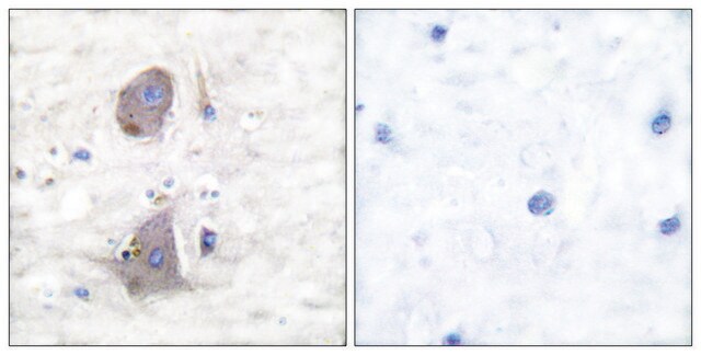
Immunocytochemistry Analysis: A representative lot detected PKC activation-induced GLUT-1 Ser226 phosphorylation in the membrane ruffles of human umbilical vein endothelial cells (HUVECs) upon TPA (Cat. No. 500582 & 524400) treatment (Lee, E.E., et al. (2015). Mol. Cell. 58(5):845-853).
Note: Process lysate samples by warming at 50°C for 10 minutes prior to gel loading. Avoid heating samples at a temperature higher than 60°C, which can cause aggregation of the target protein.
Jakość
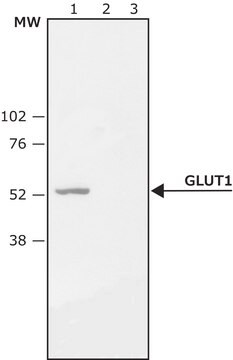
Western Blotting Analysis: 2.0 µg/mL of this antibody detected Ser226 phosphorylated GLUT-1 in lysates from PMA-treated HeLa cells.
Opis wartości docelowych
Inne uwagi
Nie możesz znaleźć właściwego produktu?
Wypróbuj nasz Narzędzie selektora produktów.
Kod klasy składowania
12 - Non Combustible Liquids
Klasa zagrożenia wodnego (WGK)
WGK 1
Temperatura zapłonu (°F)
Not applicable
Temperatura zapłonu (°C)
Not applicable
Certyfikaty analizy (CoA)
Poszukaj Certyfikaty analizy (CoA), wpisując numer partii/serii produktów. Numery serii i partii można znaleźć na etykiecie produktu po słowach „seria” lub „partia”.
Masz już ten produkt?
Dokumenty związane z niedawno zakupionymi produktami zostały zamieszczone w Bibliotece dokumentów.
Nasz zespół naukowców ma doświadczenie we wszystkich obszarach badań, w tym w naukach przyrodniczych, materiałoznawstwie, syntezie chemicznej, chromatografii, analityce i wielu innych dziedzinach.
Skontaktuj się z zespołem ds. pomocy technicznej