Przejdź do zawartości
Merck
Wszystkie zdjęcia(3)

Key Documents

07-424

Sigma-Aldrich

Anti-phospho-Histone H3 (Thr3) Antibody

Upstate®, from rabbit

Synonim(y):

H3T3P, Histone H3 (phospho T3)

Zaloguj sięWyświetlanie cen organizacyjnych i kontraktowych


About This Item

Kod UNSPSC:
12352203
eCl@ss:
32160702
NACRES:
NA.41

pochodzenie biologiczne

rabbit

Poziom jakości

forma przeciwciała

unpurified

rodzaj przeciwciała

primary antibodies

klon

polyclonal

reaktywność gatunkowa

mouse, human

producent / nazwa handlowa

Upstate®

metody

immunocytochemistry: suitable
inhibition assay: suitable
western blot: suitable

numer dostępu NCBI

numer dostępu UniProt

Warunki transportu

dry ice

docelowa modyfikacja potranslacyjna

phosphorylation (pThr3)

informacje o genach

human ... H3C1(8350)

Opis ogólny

Histone H3.1t (UniProt: Q16695; also known as H3/t, H3t, H3/g) is encoded by the HIST3H3 (also known as H3FT) gene (Gene ID: 8290) in human. Histones are highly conserved basic nuclear proteins that are responsible for the nucleosome structure of chromatin in eukaryotes. They play a central role in transcription regulation, DNA repair, DNA replication and chromosomal stability. Two molecules of each of the four core histones (H2A, H2B, H3, and H4) form an octamer, around which DNA is wrapped in repeating units, called nucleosomes, which limits DNA accessibility to the cellular machineries that require DNA as a template. Histone H3 features a main globular domain and a long N-terminal tail, which protrudes from the globular nucleosome core and can undergo several different types of epigenetic modifications that influence cellular processes. The N-terminal tails of histones are subject to posttranslational modifications, including phosphorylation, acetylation, and methylation, which recruit downstream regulatory factors, influence chromatin structure, and are critical determinants of transcription. Histone H3 can undergo acetylation on several lysine residues in the histone tail by histone acetyltransferases. Acetylation is generally associated with transcriptional activity and methylation of lysine and arginine residues can either activate or repress depending on the residue modified. Trimethylation of histone H3 is one of the most studied epigenetic marks. H3K4me3 modifications are reported to occur consistently at transcription start sites and H3K4me3 domain is associated with higher transcription activity and cell identity in pre-implantation development and in the process of deriving embryonic stem cells from the inner cell mass and trophoblast stem cells from the trophectoderm. Histone H3 is phosphorylated at threonine 4 (H3T3ph) by histone H3 associated protein kinase (HASPIN) during prophase and is dephosphorylated during anaphase and phosphorylation at serine 11 (H3S10ph) by Aurora kinase B is crucial for chromosome condensation and cell-cycle progression during mitosis and meiosis. (Ref.: Sharifi-Zarchi, A., et al. (2017). BMS Genomics. 18; Article 964; Liu, X., et al. (2016). Nature. 537(7621); 558-562).

Specyficzność

This rabbit polyclonal antibody detects Histone H3 phosphorylated on threonine 3.

Immunogen

KLH-conjugated linear peptide corresponding to 13 amino acids surrounding phosphothreonine 3 of human Histone H3.

Zastosowanie

Quality Control Testing

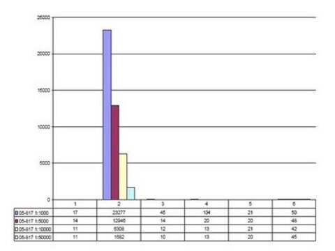
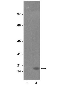
Evaluated by Western Blotting in acid extract from colcemid-treated HeLa cells.Western Blotting Analysis: A 1:5,000 dilution of this antibody detected Phospho-Histone H3 (Thr3) in acid extract of colcemid treated (50 ng/mL, overnight) HeLa cells, but not the recombinant Histone H3 protein.


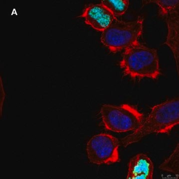
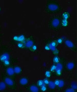
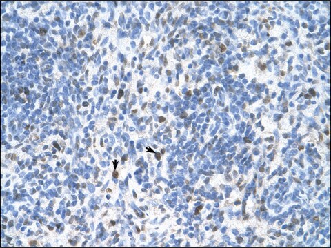
Tested ApplicationsPeptide Inhibition Assay: Target band detection in acid extract from colcemid treated (50 ng/mL; overnight) HeLa cells was prevented by preblocking of a representative lot with the immunogen phosphopeptide, but not with non-phosphorylated Histone H3 peptide.Immunocytochemistry Analysis: A 1:500 dilution from a representative lot detected Phospho-Histone H3 (Thr3) in NIH3T3 cells.Note: Actual optimal working dilutions must be determined by end user as specimens, and experimental conditions may vary with the end user.

Jakość

routinely evaluated by immunoblot in acid extracted proteins from mitotic HeLa cells

Opis wartości docelowych

~17 kDa observed; 15.51 kDa calculated. Uncharacterized bands may be observed in some lysate(s).

Postać fizyczna

Format: Unpurified
Immunodepleted
Immunodepleted rabbit serum with 0.05% sodium azide with 30% glycerol.

Przechowywanie i stabilność

Store at -10°C to -25°C. Handling Recommendations: Upon receipt and prior to removing the cap, centrifuge the vial and gently mix the solution. Aliquot into microcentrifuge tubes and store at -20°C. Avoid repeated freeze/thaw cycles, which may damage IgG and affect product performance.

Komentarz do analizy

Control
Acid extract from colcemid-treated HeLa cells

Informacje prawne

UPSTATE is a registered trademark of Merck KGaA, Darmstadt, Germany

Oświadczenie o zrzeczeniu się odpowiedzialności

Unless otherwise stated in our catalog or other company documentation accompanying the product(s), our products are intended for research use only and are not to be used for any other purpose, which includes but is not limited to, unauthorized commercial uses, in vitro diagnostic uses, ex vivo or in vivo therapeutic uses or any type of consumption or application to humans or animals.
This page may contain text that has been machine translated.

Nie możesz znaleźć właściwego produktu?  

Wypróbuj nasz Narzędzie selektora produktów.

Kod klasy składowania

12 - Non Combustible Liquids

Klasa zagrożenia wodnego (WGK)

WGK 1

Temperatura zapłonu (°F)

Not applicable

Temperatura zapłonu (°C)

Not applicable


Certyfikaty analizy (CoA)

Poszukaj Certyfikaty analizy (CoA), wpisując numer partii/serii produktów. Numery serii i partii można znaleźć na etykiecie produktu po słowach „seria” lub „partia”.

Masz już ten produkt?

Dokumenty związane z niedawno zakupionymi produktami zostały zamieszczone w Bibliotece dokumentów.

Odwiedź Bibliotekę dokumentów

AtHaspin phosphorylates histone H3 at threonine 3 during mitosis and contributes to embryonic patterning in Arabidopsis.
Raheleh Karimi Ashtiyani,Ali Mohammad Banaei Moghaddam,Veit Schubert,Twan Rutten et al.
The Plant Journal null
Yansong Wang et al.
Chembiochem : a European journal of chemical biology, 20(1), 66-71 (2018-10-20)
Protein phosphatase-1 (PP1)-disrupting peptides (PDPs) are selective chemical modulators of PP1 that liberate the active PP1 catalytic subunit from regulatory proteins; thus allowing the dephosphorylation of nearby substrates. We have optimized the original cell-active PDP3 for enhanced stability, and obtained
María Carretero et al.
The EMBO journal, 32(22), 2938-2949 (2013-10-22)
Cohesin mediates sister chromatid cohesion and contributes to the organization of interphase chromatin through DNA looping. In vertebrate somatic cells, cohesin consists of Smc1, Smc3, Rad21, and either SA1 or SA2. Three additional factors Pds5, Wapl, and Sororin bind to
Mitotic phosphorylation of histone H3 at threonine 3
Polioudaki, H., et al
Febs Letters, 560, 39-44 (2004)
Joly H L Kwek et al.
Mechanisms of development, 126(5-6), 449-463 (2009-04-17)
There are two phases of fore-stomach development during the first 200 days of pouch life in tammar wallaby. For the first 170 days, the mucosa displays an immature gastric glandular phenotype that changes to a cardia glandular phenotype, which remains

Nasz zespół naukowców ma doświadczenie we wszystkich obszarach badań, w tym w naukach przyrodniczych, materiałoznawstwie, syntezie chemicznej, chromatografii, analityce i wielu innych dziedzinach.

Skontaktuj się z zespołem ds. pomocy technicznej