추천 제품
생물학적 소스
mouse
Quality Level
결합
peroxidase conjugate
항체 형태
purified from hybridoma cell culture
항체 생산 유형
primary antibodies
클론
9E10, monoclonal
형태
lyophilized powder
종 반응성
human
포장
vial of 100 μL
농도
~2 mg/mL
기술
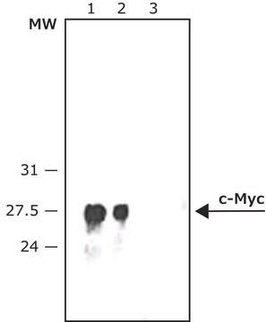
immunoblotting: 1:250-1:500 using lysate of HEK-293T cells over expressing c-Myc fusion protein
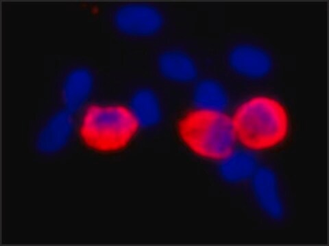
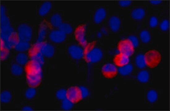
immunohistochemistry: suitable
동형
IgG1
UniProt 수납 번호
배송 상태
dry ice
저장 온도
−20°C
타겟 번역 후 변형
unmodified
유전자 정보
human ... MYC(4609)
일반 설명
Monoclonal Anti-c-Myc (mouse IgG1 isotype) is derived from the 9E10 hybridoma, produced by the fusion of mouse myeloma cells and splenocytes from a BALB/c mouse immunized with synthetic peptide of the human p62c-myc protein, conjugated to KLH. c-Myc is a proto-oncogene localized on long arm of human chromosome 8. The encoded protein contains an N-terminal transactivation domain. The human c-Myc proto-oncogene is the cellular homolog of the avian v-Myc gene found in several leukemogenic retroviruses.
특이성
Monoclonal Anti-Calponin specifically recognizes smooth muscle Calponin.
면역원
Synthetic peptide of the human p62c-myc protein.
애플리케이션
Anti-c-Myc-Peroxidase antibody, Mouse monoclonal may be used in immunoblotting and immunohistology.
생화학적/생리학적 작용
c-Myc is a transcription factor involved in regulation of cell growth, proliferation, differentiation and apoptosis. It is implicated in DNA replication, RNA splicing or transcription. Increased expression of the gene is associated with the development of various types of human cancers.
물리적 형태
Supplied as lyophilized powder. After reconstitution with 0.1 mL of distilled water to a final antibody concentration of ∼ 2 mg/mL, the solution contains 1% BSA, 2.5% trehalose, 0.05% MIT in 0.01 M sodium phosphate buffered saline
저장 및 안정성
Store the lyophilized product at 2–8 °C. For extended storage after reconstitution, keep at –20 °C in working aliquots. Avoid repeated freeze-thaw cycles. For continuous use after reconstitution, keep at 2–8 °C for up to 1 month. Solutions at working dilution should be discarded if not used within 12 hours.
면책조항
Unless otherwise stated in our catalog, our products are intended for research use only and are not to be used for any other purpose, which includes but is not limited to, unauthorized commercial uses, in vitro diagnostic uses, ex vivo or in vivo therapeutic uses or any type of consumption or application to humans or animals.
적합한 제품을 찾을 수 없으신가요?
당사의 제품 선택기 도구.을(를) 시도해 보세요.
신호어
Warning
유해 및 위험 성명서
Hazard Classifications
Skin Sens. 1
Storage Class Code
13 - Non Combustible Solids
WGK
WGK 2
Flash Point (°F)
Not applicable
Flash Point (°C)
Not applicable
시험 성적서(COA)
제품의 로트/배치 번호를 입력하여 시험 성적서(COA)을 검색하십시오. 로트 및 배치 번호는 제품 라벨에 있는 ‘로트’ 또는 ‘배치’라는 용어 뒤에서 찾을 수 있습니다.
이미 열람한 고객
K Alitalo et al.
Nature, 306(5940), 274-277 (1983-11-17)
The myelocytomatosis viruses are a family of replication-defective avian retroviruses that cause a variety of tumours in chickens and transform both fibroblasts and macrophages in culture through the activity of their oncogene v-myc. A closely related gene (c-myc) is found
S Pelengaris et al.
Current opinion in genetics & development, 10(1), 100-105 (2000-02-19)
The protein products of many dominant oncogenes are capable of inducing both cell proliferation and apoptosis. Recent experiments employing transgenic mice that express an ectopically regulatable myc gene or protein have begun to elucidate the role of the balance between
Q Hu et al.
Science (New York, N.Y.), 268(5207), 100-102 (1995-04-07)
Phosphatidylinositol (Pl)-3 kinase is one of many enzymes stimulated by growth factors. A constitutively activated mutant, p110, that functions independently of growth factor stimulation was constructed to determine the specific responses regulated by Pl-3 kinase. The p110 protein exhibited high
Ann-Marie Baker et al.
Histopathology, 69(2), 222-229 (2016-01-31)
Recent attempts to study MYC distribution in human samples have been confounded by a lack of agreement in immunohistochemical staining between antibodies targeting the N-terminus and those targeting the C-terminus of the MYC protein. The aim of this study was
D Robertson et al.
The journal of histochemistry and cytochemistry : official journal of the Histochemistry Society, 43(5), 471-480 (1995-05-01)
To determine the ultrastructural distribution of H-ras, the rho proteins rho-A, rho-B, rho-C, and the rac1 protein (members of the ras GTP-binding protein family), we used cDNA expression plasmids in which a short sequence coding for the epitope recognized by
자사의 과학자팀은 생명 과학, 재료 과학, 화학 합성, 크로마토그래피, 분석 및 기타 많은 영역을 포함한 모든 과학 분야에 경험이 있습니다..
고객지원팀으로 연락바랍니다.


