
추천 제품


생물학적 소스
mouse
Quality Level
항체 형태
purified immunoglobulin
항체 생산 유형
primary antibodies
클론
E4 (5F4D2), monoclonal
종 반응성
human, mouse, rat
기술
immunocytochemistry: suitable
immunohistochemistry: suitable (paraffin)
immunoprecipitation (IP): suitable
western blot: suitable
동형
IgG1κ
NCBI 수납 번호
UniProt 수납 번호
배송 상태
wet ice
타겟 번역 후 변형
unmodified
유전자 정보
human ... PKD1(5310)
일반 설명
Polycystin-1 (UniProt P98161; also known as Autosomal dominant polycystic kidney disease 1 protein) is encoded by the PKD1 gene (Gene ID 5310) in human. The primary cilium is a microtubule-based mechano- and chemo-sensory organelle that coordinates an array of cellular pathways during development and in tissue homeostasis, including Hedgehog (Hh), PDGFRα and Wnt signaling. Defective ciliary function causes a variety of ciliopathies, including autosomal dominant polycystic kidney disease (ADPKD). Polycystin-1 (PC1) and polycystin-2 (PC2) are large transmembrane proteins co-localized to primary cilium, where they play an important role in calcium-based signalling. PC1 is an atypical adhesion G-protein-coupled receptor (aGPCR) with 11-transmemrbane domains. PC1 is cis-autoproteolytically cleaved at a juxtamembrane GPCR cleavage site (GPS; a.a. 3012-3061) in a developmental stage-dependent manner, where PC1 is largely uncleaved in early embryonic kidneys but becomes extensively cleaved after birth. GPS cleavage results in a heterodimeric PC1 form, in which the N-terminal fragment (NTF) remains non-covalently associated with the transmembrane C-terminal fragment (CTF). Transgenic mice expressing non-cleavable PC1 develop cystic kidney disease during the postnatal period.
특이성
Clone E4 (also known as clone 5F4D2) targets polycystin-1 extracellular C-type lectin domain and immunostained wild-type, but not Pkd1-knockout murine cells (Kim, H., et al. (2014). Nat. Commun. 5:5482).
면역원
Epitope: C-type lectin domain.
FLAG-tagged recombinant human polycystin-1 C-type lectin domain.
애플리케이션
Anti-Polycystin-1 Antibody, clone E4 (5F4D2) is an antibody against Polycystin-1 for use in Immunohistochemistry (Paraffin), Immunoprecipitation, Western Blotting, Immunocytochemistry.
Immunohistochemistry Analysis: An 1:50 dilution of this antibody from a representative lot detected polycystin-1 immunoreactivity in rat and mouse kidney tissue sections.
Immunoprecipitation Analysis: A representative lot immunoprecipitated polycystin-1 from mouse lung tissue lysates (Courtesy of Dr. Feng Qian, University of Maryland School of Medicine, Baltimore, MD).
Western Blotting Analysis: A representative lot detected exogenously expressed polycystin-1 in lysates from HEK cells transfected with human or mouse Pkd1 cDNA, but not in lysates from mock-transfected HEK cells (Courtesy of Dr. Feng Qian, University of Maryland School of Medicine, Baltimore, MD).
Western Blotting Analysis: A representative lot detected the polycystin-1 (PC1) GPS domain cis-autocleaved N-terminal fragment (NTF) in lysates from HEK cells transfected with full-length Pkd1 cDNA, but not in lysates from mock-transfected HEK cells (Kim, H., et al. (2014). Nat. Commun. 5:5482).
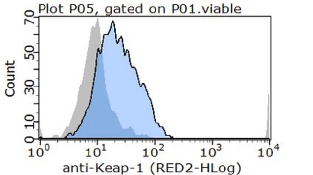
Immunocytochemistry Analysis: A representative lot and an anti-Arl13b antibody co-stained the primary cilium of 4% formaldehyde-fixed, 0.1% Triton X-100-permeabilized murine embryonic fibrolasts (mMEF) and collecting duct (CD)-derived cells by dual fluorescence immunocytochemistry. No polycystin-1 immunoreactivity was detected in Pkd1-knockout mMEFs (Kim, H., et al. (2014). Nat. Commun. 5:5482).
Immunoprecipitation Analysis: A representative lot immunoprecipitated polycystin-1 from mouse lung tissue lysates (Courtesy of Dr. Feng Qian, University of Maryland School of Medicine, Baltimore, MD).
Western Blotting Analysis: A representative lot detected exogenously expressed polycystin-1 in lysates from HEK cells transfected with human or mouse Pkd1 cDNA, but not in lysates from mock-transfected HEK cells (Courtesy of Dr. Feng Qian, University of Maryland School of Medicine, Baltimore, MD).
Western Blotting Analysis: A representative lot detected the polycystin-1 (PC1) GPS domain cis-autocleaved N-terminal fragment (NTF) in lysates from HEK cells transfected with full-length Pkd1 cDNA, but not in lysates from mock-transfected HEK cells (Kim, H., et al. (2014). Nat. Commun. 5:5482).
Immunocytochemistry Analysis: A representative lot and an anti-Arl13b antibody co-stained the primary cilium of 4% formaldehyde-fixed, 0.1% Triton X-100-permeabilized murine embryonic fibrolasts (mMEF) and collecting duct (CD)-derived cells by dual fluorescence immunocytochemistry. No polycystin-1 immunoreactivity was detected in Pkd1-knockout mMEFs (Kim, H., et al. (2014). Nat. Commun. 5:5482).
Research Category
Signaling
Signaling
Research Sub Category
Developmental Signaling
Developmental Signaling
품질
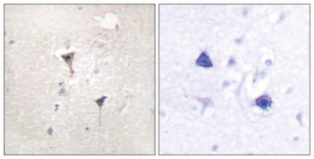
Evaluated by Immunohistochemistry in human kidney tissue.
Immunohistochemistry Analysis: An 1:250 dilution of this antibody lot detected polycystin-1 immunoreactivity in human kidney tissue.
Immunohistochemistry Analysis: An 1:250 dilution of this antibody lot detected polycystin-1 immunoreactivity in human kidney tissue.
표적 설명
Calculated molecular weights of full-length/GPS domain autocleaved N-terminal fragment: 460.3/325.0 kDa (isoform 1), 459.1/324.0 kDa (isoform 2), 460.2/325.0 kDa (isoform 3).
물리적 형태
Format: Purified
Protein G Purified
Purified mouse monoclonal IgG1κ antibody in buffer containing 0.1 M Tris-Glycine (pH 7.4), 150 mM NaCl with 0.05% sodium azide.
저장 및 안정성
Stable for 1 year at 2-8°C from date of receipt.
기타 정보
Concentration: Please refer to lot specific datasheet.
면책조항
Unless otherwise stated in our catalog or other company documentation accompanying the product(s), our products are intended for research use only and are not to be used for any other purpose, which includes but is not limited to, unauthorized commercial uses, in vitro diagnostic uses, ex vivo or in vivo therapeutic uses or any type of consumption or application to humans or animals.
적합한 제품을 찾을 수 없으신가요?
당사의 제품 선택기 도구.을(를) 시도해 보세요.
Storage Class Code
12 - Non Combustible Liquids
WGK
WGK 1
Flash Point (°F)
Not applicable
Flash Point (°C)
Not applicable
시험 성적서(COA)
제품의 로트/배치 번호를 입력하여 시험 성적서(COA)을 검색하십시오. 로트 및 배치 번호는 제품 라벨에 있는 ‘로트’ 또는 ‘배치’라는 용어 뒤에서 찾을 수 있습니다.
자사의 과학자팀은 생명 과학, 재료 과학, 화학 합성, 크로마토그래피, 분석 및 기타 많은 영역을 포함한 모든 과학 분야에 경험이 있습니다..
고객지원팀으로 연락바랍니다.