コンテンツへスキップ
Merck
ホームタンパク質発現上皮成長因子受容体 (EGFR) シグナリング

上皮成長因子受容体 (EGFR) シグナリング

背景
上皮成長因子受容体 (EGFR) ・リガンド
    受容体活性化
EGFR シグナリング
    Ras/Raf シグナリングカスケード
    ホスファチジルイノシトール3キナーゼ/Akt シグナリングカスケード
    シグナル伝達性転写因子 (STAT) 経路
    ホスホリパーゼCγシグナリング
    Nck/PAKシグナリングカスケード
    Cbl仲介型エンドサイトーシス
    EGFRの核内への移動
分化中・疾患時のEGFR
まとめ

背景

上皮成長因子受容体(EGFR)は受容体チロシンキナーゼファミリー(RTK)に属し、ファミリーの中で最初に発見されたメンバーです。細胞外環境でさまざまなリガンドによって活性化され、細胞反応を伝達し、細胞増殖、細胞生存、成長や分化など、さまざまな細胞活動に介在します。EGFRは多くの臓器で発現しており、異常な発現はさまざまながんに関与しています。本レビューは主として、EGFR経路のさまざまなシグナリングとそのシグナル伝達経路について紹介します。

上皮成長因子受容体 (EGFR)とリガンド

ErbBファミリーはEGFR(ErbB-1/HER1)、ErbB-2(Neu、HER2)、ErbB-3(HER3)、およびErbB-4(HER4)という4つの受容体で構成されます。このタンパク質の受容体チロシンキナーゼファミリー(RTK)には細胞外リガンド結合ドメイン、疎水性膜貫通ドメイン、および細胞質チロシンキナーゼドメインがあります (表1)。ErbB受容体は、結合特異性を生じさせる3つのジスルフィド結合が特徴的なEGFファミリーの成長因子によって活性化されます。さらに構造的モチーフとして、免疫グロブリン様ドメイン、ヘパリン結合部位、およびグリコシル化部位があります1,2。 
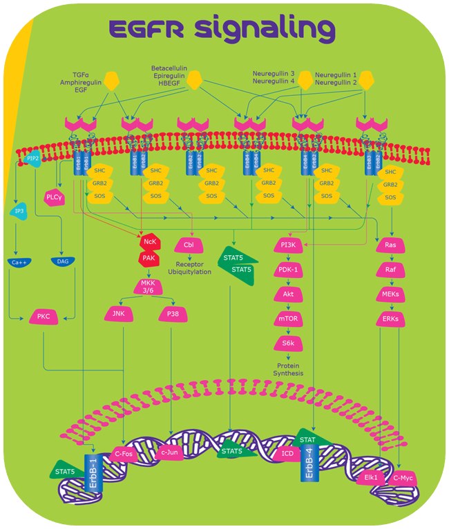
表 1シグナル受容体・リガンド

受容体活性化

受容体活性化は細胞膜上でのカスケード反応が引き金になります。

  • リガンド結合各リガンドが同族ErbB受容体の細胞外ドメインに結合します
  • 受容体の二量体化リガンド結合によって受容体のホモまたはヘテロ二量体形成が誘発されます3
  • キナーゼドメインの活性化カルボキシ基末端の主要チロシン残基が自己リン酸化されて受容体を活性化し、タンパク質がSrcホモロジー2(SH2)およびホスホチロシン結合ドメイン(PTB)にドッキングする部位として働き、細胞内シグナリングの引き金になります3,4。 
EGFRシグナリング

図1.EGFRシグナリング

EGFRシグナリング

Ras/Rafシグナリングカスケード

受容体:2つのErbB受容体の二量体

主な機能:細胞生存・細胞増殖5

ErbB受容体の活性化後、Grb2およびSosから成る複合体が直接またはアダプタータンパク質Shcの関与により、受容体の特異的チロシン残基に結合します6,7。これによりSosの立体構造が変化し、Ras-GDPをリクルートし活性化することが可能になります。Ras-GDPはRaf-1を活性化し、これによりマイトジェン活性化タンパク質キナーゼ(MAPK)を介してさらに細胞外シグナル調節キナーゼ1および2(ERK1およびERK2)を活性化します8,9。活性化されたキナーゼは最終的に核内に移動し、Elk1やC-mycなどの特異的転写因子をリン酸化し、細胞増殖を誘導します。

ホスファチジルイノシトール3キナーゼ/Akt シグナリングカスケード

受容体:ErbB4またはErbB3とErbB2との二量体

主な機能:細胞成長、アポトーシス抵抗、細胞浸潤・遊走10

ホスファチジルイノシトールはp85およびp110サブユニットから成ります。2つのサブユニットはErbB受容体に結合し、二次伝達物質であるホスファチジルイノシトール3,4,5-三リン酸を生成し、さらにセリン/スレオニンキナーゼAKTを活性化します。この活性化により、AKTがmTORをリン酸化し、続いてS6Kがリン酸化されることでタンパク質合成に至ります10

シグナル伝達性転写因子 (STAT) 経路

受容体:ErbB

主な機能:腫瘍の進行、腫瘍発生、および血管新生11

STATタンパク質がSrcホモロジー2ドメインを介してErbB受容体のリン酸化チロシン残基に結合し、二量体化が起きると核内に移動し、Myc、Nos2、p21、サイトカインなどの特異的標的遺伝子の発現を促します12

ホスホリパーゼCγシグナリング

受容体ErbB1

主な機能:イオンチャネル、細胞遊走、カルシウム仲介型シグナリングの調節13

ホスホリパーゼCγはErbB1と相互作用を起こし、ホスファチジルイノシトール4,5-二リン酸(PIP2)を加水分解してイノシトール1,3,5-三リン酸(IP3)および1,2-ジアシルグリセロール(DAG)を生成します。IP3は細胞内カルシウム濃度を上昇させ、DAGはタンパク質キナーゼC(PKC)の活性化を仲介します13。  次に活性化されたPKCがMAPKおよびc-Jun NH2末端キナーゼを活性化します14

Nck/PAKシグナリングカスケード

受容体:ErbB1

主な機能:細胞生存・細胞遊走15

NckはSH2ドメインを含むアダプタータンパク質で、SH2ドメインはEGF受容体に結合し、下流のシグナリングの引き金となります。NckはSH3ドメインを介してPAK1(p21/CDC42/Rac1活性化キナーゼ-1)の結合を活性化します。次に活性化されたPAK1がMEKK1(MAP/ERKキナーゼ-1)およびMKK4/7(MAPキナーゼ-4/7)を介してJNK(c-Junキナーゼ)をそれぞれ活性化します。JNKは核内に移動し、c-Fosおよびc-Junなどの転写因子をリン酸化します16

Cbl仲介型エンドサイトーシス

受容体:ErbB1

主な機能:エンドサイトーシス

リガンド結合後、CblはSH2ドメインまたはGRB2アダプタータンパク質を介してEGF受容体に結合する基質になり、受容体のリソソーム分解の引き金となります17

EGFRの核内への移動

EGF受容体にはリソソーム分解を逃れて核内に移動し、生物機能を調節します。核内でEGF受容体は Cyclin D1 遺伝子などの細胞生死遺伝子の転写を促し、さらにSTATおよびE2F1転写因子の補助因子として働きます18。EGFRの核局在化により抗腫瘍mAbの増殖抑制作用に対する抵抗性が生じ、疾患重症度に関わってきます19

メルクの高品質で人気の高いEGFタンパク質を研究にご利用ください

分化中・疾患時のEGFR

EGF受容体およびそのリガンドはさまざまな臓器の分化に重要です。EGFRノックアウトモデルは胚致死性で組織/臓器が不完全になります。しかし、ErbBリガンドの変異は致死性の表現型ではありません。他のリガンドの補助経路が、ErbBリガンド変異によって失われたシグナルを補ってくれるためです。20-25多くの疾患ではEGFRが異常に活性化・発現しています。特に、がん進行におけるEGFRの役割については研究が進んでいます(表 2)。 

表2ヒトの病態におけるEGFR受容体の遺伝的変化

結論

EGFRシグナリングは発生において多面的に機能し、さまざまな生理機能を調節します。各種病態にEGF受容体の変異発現が関与していると言われるようになりました。脳、心臓、皮膚、腎臓、乳腺、肺など、さまざまな臓器の発生におけるEGFRシグナリングの役割を詳細に解明するには、いまださらなる研究が必要です。薬剤抵抗性がんと闘うために、有効性が高く、薬物活性が持続し、交差反応が少ない新規薬剤を見出す試みや研究は今後も大いなる関心を呼ぶでしょう。

1.
Arteaga C, Engelman J. 2014. ERBB Receptors: From Oncogene Discovery to Basic Science to Mechanism-Based Cancer Therapeutics. Cancer Cell. 25(3):282-303. https://doi.org/10.1016/j.ccr.2014.02.025
2.
Kovacs E, Zorn JA, Huang Y, Barros T, Kuriyan J. 2015. A Structural Perspective on the Regulation of the Epidermal Growth Factor Receptor. Annu. Rev. Biochem.. 84(1):739-764. https://doi.org/10.1146/annurev-biochem-060614-034402
3.
Capuani F, Conte A, Argenzio E, Marchetti L, Priami C, Polo S, Di Fiore PP, Sigismund S, Ciliberto A. 2015. Quantitative analysis reveals how EGFR activation and downregulation are coupled in normal but not in cancer cells. Nat Commun. 6(1): https://doi.org/10.1038/ncomms8999
4.
Zhang X, Gureasko J, Shen K, Cole PA, Kuriyan J. 2006. An Allosteric Mechanism for Activation of the Kinase Domain of Epidermal Growth Factor Receptor. Cell. 125(6):1137-1149. https://doi.org/10.1016/j.cell.2006.05.013
5.
Gaestel M. 2006. MAPKAP kinases ? MKs ? two's company, three's a crowd. Nat Rev Mol Cell Biol. 7(2):120-130. https://doi.org/10.1038/nrm1834
6.
Lowenstein E, Daly R, Batzer A, Li W, Margolis B, Lammers R, Ullrich A, Skolnik E, Bar-Sagi D, Schlessinger J. 1992. The SH2 and SH3 domain-containing protein GRB2 links receptor tyrosine kinases to ras signaling. Cell. 70(3):431-442. https://doi.org/10.1016/0092-8674(92)90167-b
7.
Batzer AG, Rotin D, Ureña JM, Skolnik EY, Schlessinger J. 1994. Hierarchy of binding sites for Grb2 and Shc on the epidermal growth factor receptor.. Mol. Cell. Biol.. 14(8):5192-5201. https://doi.org/10.1128/mcb.14.8.5192
8.
B H, S I R, J D. 1994. Interaction of Ras and Raf in intact mammalian cells upon extracellular stimulation. J Biol Chem. 269(6):3913-6.
9.
Liebmann C. 2001. Regulation of MAP kinase activity by peptide receptor signalling pathway: Paradigms of multiplicity. Cellular Signalling. 13(11):777-785. https://doi.org/10.1016/s0898-6568(01)00192-9
10.
Vivanco I, Sawyers CL. 2002. The phosphatidylinositol 3-Kinase?AKT pathway in human cancer. Nat Rev Cancer. 2(7):489-501. https://doi.org/10.1038/nrc839
11.
Niu G, Wright KL, Huang M, Song L, Haura E, Turkson J, Zhang S, Wang T, Sinibaldi D, Coppola D, et al. 2002. Constitutive Stat3 activity up-regulates VEGF expression and tumor angiogenesis. Oncogene. 21(13):2000-2008. https://doi.org/10.1038/sj.onc.1205260
12.
Bromberg J. 2002. Stat proteins and oncogenesis. J. Clin. Invest.. 109(9):1139-1142. https://doi.org/10.1172/jci0215617
13.
Patterson RL, van Rossum DB, Nikolaidis N, Gill DL, Snyder SH. 2005. Phospholipase C-?: diverse roles in receptor-mediated calcium signaling. Trends in Biochemical Sciences. 30(12):688-697. https://doi.org/10.1016/j.tibs.2005.10.005
14.
Scho?nwasser DC, Marais RM, Marshall CJ, Parker PJ. 1998. Activation of the Mitogen-Activated Protein Kinase/Extracellular Signal-Regulated Kinase Pathway by Conventional, Novel, and Atypical Protein Kinase C Isotypes. Mol. Cell. Biol.. 18(2):790-798. https://doi.org/10.1128/mcb.18.2.790
15.
Ye DZ, Field J. 2012. PAK signaling in cancer. Cellular Logistics. 2(2):105-116. https://doi.org/10.4161/cl.21882
16.
Tomar A, Schlaepfer DD. 2010. A PAK-Activated Linker for EGFR and FAK. Developmental Cell. 18(2):170-172. https://doi.org/10.1016/j.devcel.2010.01.013
17.
Yu P, Fan Y, Qu X, Zhang J, Song N, Liu J, Liu Y. 2016. Cbl-b regulates the sensitivity of cetuximab through ubiquitin-proteasome system in human gastric cancer cells. J Buon. 21(4):867-873.
18.
Lo H, Hsu S, Ali-Seyed M, Gunduz M, Xia W, Wei Y, Bartholomeusz G, Shih J, Hung M. 2005. Nuclear interaction of EGFR and STAT3 in the activation of the iNOS/NO pathway. Cancer Cell. 7(6):575-589. https://doi.org/10.1016/j.ccr.2005.05.007
19.
Lo H, Xia W, Wei Y, MA, Huang S, Hung M. 2005. Novel prognostic value of nuclear epidermal growth factor receptor in breast cancer. Cancer Res. 65(1):338-48.
20.
Miettinen PJ, Berger JE, Meneses J, Phung Y, Pedersen RA, Werb Z, Derynck R. 1995. Epithelial immaturity and multiorgan failure in mice lacking epidermal growth factor receptor. Nature. 376(6538):337-341. https://doi.org/10.1038/376337a0
21.
Sibilia M, Wagner E. 1995. Strain-dependent epithelial defects in mice lacking the EGF receptor. Science. 269(5221):234-238. https://doi.org/10.1126/science.7618085
22.
Park S, Miller R, Krane I, Vartanian T. 2001. The erbB2 gene is required for the development of terminally differentiated spinal cord oligodendrocytes. 154(6):1245-1258. https://doi.org/10.1083/jcb.200104025
23.
Leu M. 2003. Erbb2 regulates neuromuscular synapse formation and is essential for muscle spindle development. 130(11):2291-2301. https://doi.org/10.1242/dev.00447
24.
S L E, K S O, N G, L L, G F, M B, L H L, M W M. 1997. ErbB3 is required for normal cerebellar and cardiac development: a comparison with ErbB2-and heregulin-deficient mice. Development. 124(24):4999-5011.
25.
Golub MS, Germann SL, Lloyd K. 2004. Behavioral characteristics of a nervous system-specific erbB4 knock-out mouse. Behavioural Brain Research. 153(1):159-170. https://doi.org/10.1016/j.bbr.2003.11.010
26.
Maheswaran S, Sequist LV, Nagrath S, Ulkus L, Brannigan B, Collura CV, Inserra E, Diederichs S, Iafrate AJ, Bell DW, et al. 2008. Detection of Mutations inEGFRin Circulating Lung-Cancer Cells. N Engl J Med. 359(4):366-377. https://doi.org/10.1056/nejmoa0800668
27.
Montagut C, Dalmases A, Bellosillo B, Crespo M, Pairet S, Iglesias M, Salido M, Gallen M, Marsters S, Tsai SP, et al. 2012. Identification of a mutation in the extracellular domain of the Epidermal Growth Factor Receptor conferring cetuximab resistance in colorectal cancer. Nat Med. 18(2):221-223. https://doi.org/10.1038/nm.2609
28.
Rosell R, Moran T, Queralt C, Porta R, Cardenal F, Camps C, Majem M, Lopez-Vivanco G, Isla D, Provencio M, et al. 2009. Screening for Epidermal Growth Factor Receptor Mutations in Lung Cancer. N Engl J Med. 361(10):958-967. https://doi.org/10.1056/nejmoa0904554
29.
Liu Z, Hou P, Ji M, Guan H, Studeman K, Jensen K, Vasko V, El-Naggar AK, Xing M. 2008. Highly Prevalent Genetic Alterations in Receptor Tyrosine Kinases and Phosphatidylinositol 3-Kinase/Akt and Mitogen-Activated Protein Kinase Pathways in Anaplastic and Follicular Thyroid Cancers. The Journal of Clinical Endocrinology & Metabolism. 93(8):3106-3116. https://doi.org/10.1210/jc.2008-0273
30.
Gao SP, Mark KG, Leslie K, Pao W, Motoi N, Gerald WL, Travis WD, Bornmann W, Veach D, Clarkson B, et al. 2007. Mutations in the EGFR kinase domain mediate STAT3 activation via IL-6 production in human lung adenocarcinomas. J. Clin. Invest.. 117(12):3846-3856. https://doi.org/10.1172/jci31871
31.
Fukushige S, Murotsu T, Matsubara K. 1986. Chromosomal assignment of human genes for gastrin, thyrotropin (TSH)-? subunit and C-erbB-2 by chromosome sorting combined with velocity sedimentation and southern hybridization. Biochemical and Biophysical Research Communications. 134(2):477-483. https://doi.org/10.1016/s0006-291x(86)80445-4
32.
Slamon D, Godolphin W, Jones L, Holt J, Wong S, Keith D, Levin W, Stuart S, Udove J, Ullrich A, et al. 1989. Studies of the HER-2/neu proto-oncogene in human breast and ovarian cancer. Science. 244(4905):707-712. https://doi.org/10.1126/science.2470152
33.
Qiu Y, Ravi L, Kung H. 1998. Requirement of ErbB2 for signalling by interleukin-6 in prostate carcinoma cells. Nature. 393(6680):83-85. https://doi.org/10.1038/30012
34.
Harskamp LR, Gansevoort RT, van Goor H, Meijer E. 2016. The epidermal growth factor receptor pathway in chronic kidney diseases. Nat Rev Nephrol. 12(8):496-506. https://doi.org/10.1038/nrneph.2016.91
35.
Scafidi J, Hammond TR, Scafidi S, Ritter J, Jablonska B, Roncal M, Szigeti-Buck K, Coman D, Huang Y, McCarter RJ, et al. 2014. Intranasal epidermal growth factor treatment rescues neonatal brain injury. Nature. 506(7487):230-234. https://doi.org/10.1038/nature12880
36.
Campbell P, Morton PE, Takeichi T, Salam A, Roberts N, Proudfoot LE, Mellerio JE, Aminu K, Wellington C, Patil SN, et al. 2014. Epithelial Inflammation Resulting from an Inherited Loss-of-Function Mutation in EGFR. Journal of Investigative Dermatology. 134(10):2570-2578. https://doi.org/10.1038/jid.2014.164
ログインして続行

続きを確認するには、ログインするか、新規登録が必要です。

アカウントをお持ちではありませんか?