
おすすめの製品

由来生物
rabbit
結合体
unconjugated
抗体製品の状態
affinity isolated antibody
抗体製品タイプ
primary antibodies
クローン
polyclonal
製品種目
Prestige Antibodies® Powered by Atlas Antibodies
フォーム
buffered aqueous glycerol solution
交差性
human
強化検証
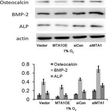
RNAi knockdown
orthogonal RNAseq
Learn more about Antibody Enhanced Validation
テクニック
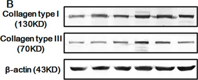
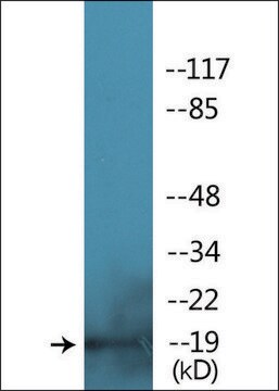
immunoblotting: 0.04-0.4 μg/mL
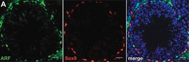
immunofluorescence: 0.25-2 μg/mL
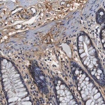
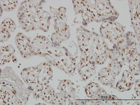
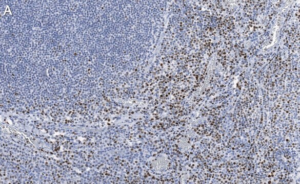
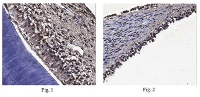
immunohistochemistry: 1:200-1:500
免疫原配列
LNSAPSPFNPQGQSQITDPRQAQSSPPWSYDQSYPSYLSQMTSPSIHSTTPLSSTRGTGLPAITDVPRRISGASELGPFSDPRQFPSISSLTESRFSNPRMHYPA
UniProtアクセッション番号
アプリケーション
research pathology
輸送温度
wet ice
保管温度
−20°C
ターゲットの翻訳後修飾
unmodified
遺伝子情報
human ... RUNX2(860)
詳細
特異性
免疫原
アプリケーション
The Human Protein Atlas project can be subdivided into three efforts: Human Tissue Atlas, Cancer Atlas, and Human Cell Atlas. The antibodies that have been generated in support of the Tissue and Cancer Atlas projects have been tested by immunohistochemistry against hundreds of normal and disease tissues and through the recent efforts of the Human Cell Atlas project, many have been characterized by immunofluorescence to map the human proteome not only at the tissue level but now at the subcellular level. These images and the collection of this vast data set can be viewed on the Human Protein Atlas (HPA) site by clicking on the Image Gallery link. We also provide Prestige Antibodies® protocols and other useful information.
生物化学的/生理学的作用
特徴および利点
Every Prestige Antibody is tested in the following ways:
- IHC tissue array of 44 normal human tissues and 20 of the most common cancer type tissues.
- Protein array of 364 human recombinant protein fragments.
関連事項
物理的形状
法的情報
免責事項
適切な製品が見つかりませんか。
製品選択ツール.をお試しください
保管分類コード
10 - Combustible liquids
WGK
WGK 1
引火点(°F)
Not applicable
引火点(℃)
Not applicable
最新バージョンのいずれかを選択してください:
資料
Mesenchymal stem cell markers and antibodies suitable for investigating targets in fibroblasts, chondrocytes, adipocytes, osteoblasts, and muscle cells.
線維芽細胞、軟骨細胞、脂肪細胞、骨芽細胞、および筋細胞のターゲットの研究に適した間葉系幹細胞マーカーおよび抗体。
ライフサイエンス、有機合成、材料科学、クロマトグラフィー、分析など、あらゆる分野の研究に経験のあるメンバーがおります。.
製品に関するお問い合わせはこちら(テクニカルサービス)