
おすすめの製品


由来生物
mouse
品質水準
抗体製品の状態
purified immunoglobulin
抗体製品タイプ
primary antibodies
クローン
NE14, monoclonal
交差性
rat, pig, human
メーカー/製品名
Chemicon®
テクニック
immunohistochemistry: suitable
アイソタイプ
IgG1
NCBIアクセッション番号
UniProtアクセッション番号
輸送温度
wet ice
ターゲットの翻訳後修飾
unmodified
遺伝子情報
human ... NEFH(4744)
pig ... Nefh(100156492)
rat ... Nefh(24587)
詳細
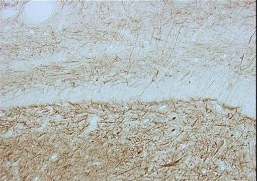
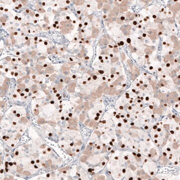
Neurofilaments are a type of intermediate filament that serve as major elements of the cytoskeleton supporting the axon cytoplasm. They are the most abundant fibrillar components of the axon, being on average 3-10 times more frequent than axonal microtubules. Neurofilaments (10nm in dia.) are built from three intertwined protofibrils which are themselves composed of two tetrameric protofilament complexs of monomeric proteins. The neurofilament triplet proteins (68/70, 160, and 200 kDa) occur in both the central and peripheral nervous system and are usually neuron specific. The 68/70 kDa NF-L protein can self-assemble into a filamentous structure, however the 160 kDa NF-M and 200 kDa NF-H proteins require the presence of the 68/70 kDa NF-L protein to co-assemble. Neuromas, ganglioneuromas, gangliogliomas, ganglioneuroblastomas and neuroblastomas stain positively for neurofilaments. Although typically restricted to neurons, neurofilaments have been detected in paragangliomas and adrenal and extra-adrenal pheochromocytomas. Carcinoids, neuroendocrine carcinomas of the skin, and oat cell carcinomas of the lung also express neurofilaments. For more neurofilament information see Nervous System Cell Type Specific Marker chart online under the CHEMICON Technical Support section.
特異性
The antibody reacts with neurofilament 200 kD.
免疫原
Purified neurofilament polypeptides (Debus et al., 1983).
アプリケーション
Research Category
ニューロサイエンス
ニューロサイエンス
Research Sub Category
ニューロフィラメント及び神経細胞代謝
神経細胞及びグリアマーカー
ニューロフィラメント及び神経細胞代謝
神経細胞及びグリアマーカー
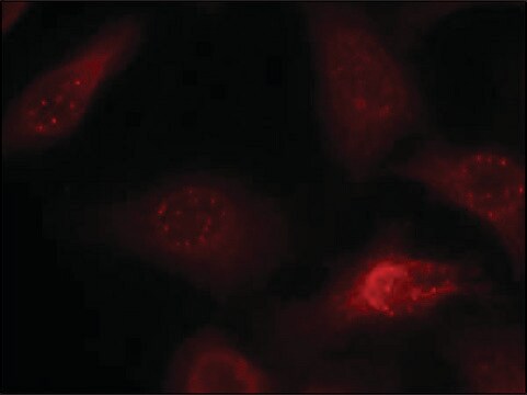
Detect Neurofilament 200 kDa using this Anti-Neurofilament 200 kDa Antibody, clone NE14 validated for use in IH.
Immunohistochemistry: 5-10 μg/mL (See below protocol.)
Optimal working dilutions must be determined by end user.
Immunohistochemistry Protocol for Anti-Neurofilament 200 kD
Ideal specimens are obtained from frozen sections from shock-frozen tissue samples. The frozen sections are dried in the air and then fixed with acetone at -15 to -25°C for 10 min. Excess acetone is allowed to evaporate at 15-25°C. Material fixed in alcohol and embedded in paraffin can also be used, see (Altmannsberger et al., 1982). The antibody appears to react with tissue fixed in formaldehyde for a short time (10 min) (Debus et al., 1983). Other fixation conditions must be first tested by the investigator.
It is advantageous to block unspecific binding sites by overlaying the sections with fetal calf serum for 20-30 min at 15-25°C. Excess of fetal calf serum is removed by decanting before application of the antibody solution. Cytocentrifuge preparations of single cells or cell smears are also fixed in acetone. These preparations should, however, not be dried in the air. Instead, the excess acetone is removed by briefly washing in phosphate-buffered saline (PBS).
Further treatment is then as follows:
Overlay the preparation with 10-20 μL antibody solution and incubate in a humid chamber at 37°C for 1 h.
Dip the slide briefly in PBS and then wash 3 times in PBS for 3 min (use fresh PBS each time)
Wipe the margins of the preparation dry and overlay the preparation with 10-20 μL of an anti-mouse Ig-FITC or anti-mouse Ig-POD antibody and allow to incubate for 1 h at 37°C in a humid chamber.
Wash the slide as described above.
The preparation must not be allowed to dry out during any of the steps.
If using an indirect immunofluorescence technique, the preparation should be overlaid with a suitable embedding medium (e.g. Moviol, Hoechst) and examined under the fluorescence microscope. If a POD-conjugate has been used as the secondary antibody, the preparation should be overlaid with a substrate solution (see below) and incubated at 15-25°C until a clearly visible red-brown color develops. A negative control (e.g. only the secondary antibody) should remain unchanged in color during this incubation period. Subsequently, the substrate is washed off with PBS and the preparation is stained, if desired, with hemalum stain for about 1 min. The hemalum solution is washed off with PBS, the preparation is embedded and examined.
Substrate solutions:
Aminoethyl-carbazole:
Dissolve 2 mg 3-amino-9-ethylcarbazole with 1.2 mL dimethylsulfoxide and add 28.8 mL 0.05 M Tris-HCl, pH 7.3, and 20 μL 3% H 2 O 2 (w/v). Prepare solution freshly each day.
Diaminobenzidine:
Dissolve 25 mg 3,3′-diaminobenzidine with 50 mL 0.05 M Tris-HCl, pH 7.3, and add 40 μL 3% H2O2 (w/v). Prepare solution freshly each day.
物理的形状
Format: Purified
Purified immunoglobulin. Liquid. Buffer = 0.02M Phosphate buffer, 0.25M NaCl containing 0.1% sodium azide.
保管および安定性
Maintain refrigerated at 2-8°C in undiluted aliquots for up to 6 months.DO NOT FREEZE
その他情報
Concentration: Please refer to the Certificate of Analysis for the lot-specific concentration.
法的情報
CHEMICON is a registered trademark of Merck KGaA, Darmstadt, Germany
免責事項
Unless otherwise stated in our catalog or other company documentation accompanying the product(s), our products are intended for research use only and are not to be used for any other purpose, which includes but is not limited to, unauthorized commercial uses, in vitro diagnostic uses, ex vivo or in vivo therapeutic uses or any type of consumption or application to humans or animals.
適切な製品が見つかりませんか。
製品選択ツール.をお試しください
保管分類コード
10 - Combustible liquids
WGK
WGK 2
引火点(°F)
Not applicable
引火点(℃)
Not applicable
試験成績書(COA)
製品のロット番号・バッチ番号を入力して、試験成績書(COA) を検索できます。ロット番号・バッチ番号は、製品ラベルに「Lot」または「Batch」に続いて記載されています。
Neurochemical characterization of insulin receptor-expressing primary sensory neurons in wild-type and vanilloid type 1 transient receptor potential receptor knockout mice.
Djalil Baiou,Peter Santha,Antonio Avelino,Ana Charrua,Timea Bacskai,Klara Matesz et al.
The Journal of Comparative Neurology null
Immunohistochemical localization of histamine receptor subtypes in human inferior turbinates.
Muneo Nakaya, Naonobu Takeuchi, Kenji Kondo
The Annals of Otology, Rhinology, and Laryngology null
Asiyeh Golabchi et al.
Biosensors & bioelectronics, 155, 112096-112096 (2020-02-25)
Intracortical microelectrodes are being developed to both record and stimulate neurons to understand brain circuitry or restore lost functions. However, the success of these probes is hampered partly due to the inflammatory host tissue responses to implants. To minimize the
Takashi D Y Kozai et al.
Biomaterials, 35(36), 9620-9634 (2014-09-02)
Chronic implantation of microelectrodes into the cortex has been shown to lead to inflammatory gliosis and neuronal loss in the microenvironment immediately surrounding the probe, a hypothesized cause of neural recording failure. Caspase-1 (aka Interleukin 1β converting enzyme) is known
Asiyeh Golabchi et al.
Biomaterials, 225, 119519-119519 (2019-10-11)
The inflammatory brain tissue response to implanted neural electrode devices has hindered the longevity of these implants. Zwitterionic polymers have a potent anti-fouling effect that decreases the foreign body response to subcutaneous implants. In this study, we developed a nanoscale
ライフサイエンス、有機合成、材料科学、クロマトグラフィー、分析など、あらゆる分野の研究に経験のあるメンバーがおります。.
製品に関するお問い合わせはこちら(テクニカルサービス)