おすすめの製品
由来生物
mouse
抗体製品の状態
purified immunoglobulin
抗体製品タイプ
primary antibodies
クローン
QBEnd/10, monoclonal
交差性
human, monkey
包装
antibody small pack of 25 μL
テクニック
electron microscopy: suitable
flow cytometry: suitable
immunofluorescence: suitable
immunohistochemistry: suitable (paraffin)
western blot: suitable
アイソタイプ
IgG1λ
NCBIアクセッション番号
UniProtアクセッション番号
ターゲットの翻訳後修飾
unmodified
遺伝子情報
human ... CD34(947)
関連するカテゴリー
詳細
Hematopoietic progenitor cell antigen CD34 (UniProt: P28906; also known as CD34) is encoded by the CD34 gene (Gene ID: 947) in human. CD34 is a highly glycosylated single-pass type I membrane protein that is expressed on hematopoietic progenitor cells and small vessel endothelium of a variety of tissues. Under normal conditions, CD34+ expressing cells account for about 1 2% of the total bone marrow cells. It serves as an adhesion molecule that plays a role in early hematopoiesis by mediating the attachment of stem cells to the bone marrow extracellular matrix or directly to stromal cells. It is also reported to act as a scaffold for the attachment of lineage specific glycans, allowing stem cells to bind to lectins expressed by stromal cells or other marrow components. CD34 is synthesized with a signal peptide (aa 1-31) that is cleaved off in the mature form. The mature form has an extracellular domain (aa 32-290), a transmembrane domain (aa 291-311), and a cytoplasmic domain (aa 312-385). Two isoforms of CD34 have been described that are produced by alternative splicing.
特異性
Clone QBEnd/10 specifically detects CD34 in human and non-human primates.
免疫原
Human placental endothelial membrane vesicles.
アプリケーション
Anti-CD34, clone QBEnd/10, Cat. No. CBL496-I, is a mouse monoclonal antibody that detects CD34 and has been tested for use in Electron Microscopy, Flow Cytometry, Immunofluorescence and Fluorescence Activated Cell Sorting (FACS), Immunohistochemistry (Paraffin), and Western Blotting.
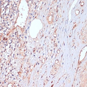
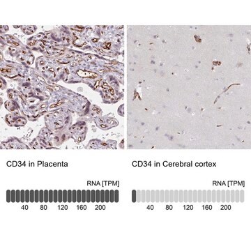
Immunohistochemistry (Paraffin) Analysis: A 1:250 dilution from a representative lot detected CD34 in human brain tissue sections.
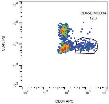
Fluorescence Activated Cell Sorting (FACS) Analysis: A representative lot was used to sort CD34+ cells from bone marrow. (de Bock, C.E., et. al. (2012). Leukemia. 26(5):918-26).
Immunofluorescence Analysis: A representative lot detected CD34 in Immunofluorescence applications (Miki, T., et. al. (2010). Mol Cancer Res. 8(5):665-76).
Electron Microscopy Analysis: A representative lot detected CD34 in Electron Microscopy applications (Fina, L., et. al. (1990). Blood. 75(12):2417-26).
Immunohistochemistry Analysis: A representative lot detected CD34 in Immunohistochemistry applications (Engler, J.R., et. al. (2012). PLoS One. 7(8):e43339; Fina, L., et. al. (1990). Blood. 75(12):2417-26).
Flow Cytometry Analysis: A representative lot detected CD34 in Flow Cytometry applications (de Bock, C.E., et. al. (2012). Leukemia. 26(5):918-26; Fina, L., et. al. (1990). Blood. 75(12):2417-26).
Western Blotting Analysis: A representative lot detected CD34 in Western Blotting applications (Fina, L., et. al. (1990). Blood. 75(12):2417-26).
品質
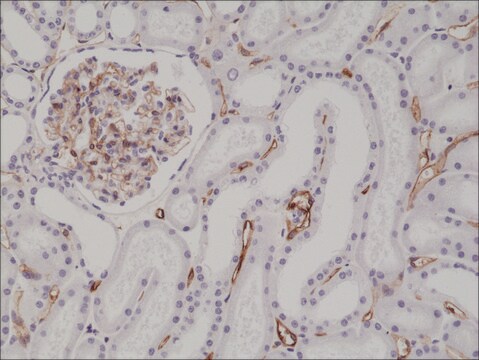
Evaluated by Immunohistochemistry (Paraffin) in human kidney tissue sections.
Immunohistochemistry (Paraffin) Analysis: A 1:250 dilution of this antibody detected CD34 in human kidney tissue sections.
Immunohistochemistry (Paraffin) Analysis: A 1:250 dilution of this antibody detected CD34 in human kidney tissue sections.
ターゲットの説明
40.72 kDa Calculated. This antibody recognizes a heavily glycosylated transmembrane protein: gp 105-120 kDa
物理的形状
Format: Purified
その他情報
Concentration: Please refer to lot specific datasheet.
適切な製品が見つかりませんか。
製品選択ツール.をお試しください
試験成績書(COA)
製品のロット番号・バッチ番号を入力して、試験成績書(COA) を検索できます。ロット番号・バッチ番号は、製品ラベルに「Lot」または「Batch」に続いて記載されています。
ライフサイエンス、有機合成、材料科学、クロマトグラフィー、分析など、あらゆる分野の研究に経験のあるメンバーがおります。.
製品に関するお問い合わせはこちら(テクニカルサービス)