
おすすめの製品


由来生物
rabbit
品質水準
抗体製品の状態
serum
抗体製品タイプ
primary antibodies
クローン
polyclonal
交差性
human
テクニック
immunocytochemistry: suitable
western blot: suitable
NCBIアクセッション番号
UniProtアクセッション番号
輸送温度
ambient
ターゲットの翻訳後修飾
unmodified
遺伝子情報
human ... CENPC(1060)
詳細
Centromere protein C (UniProt Q03188; also known as CENP-C, CENP-C 1, Centromere autoantigen C, Centromere protein C 1, Interphase centromere complex protein 7) is encoded by the CENPC (also known as CENPC1, ICEN7) gene (Gene ID 1060) in human. Kinetochores assemble on centromeres to bind spindle microtubules and mediate chromosome segregation during cell division. CENP-C is a subunit of constitutive centromere-associated network (CCAN) that acts as the centromere-kinetochore interface. CCAN is a large complex composed of at least 16 different centromeric proteins (CENPs). CENP-C plays an important role in kinetochore assembly downstream of CENP-A. The PEST domain in the N-terminal half of CENP-C interacts directly with the CCAN subcomplex CENP-HIKM (CENP-H, CENP-K, CENP-I, and CENP-M) and CENP-C depletion is shown to cause mislocalization of CENP-T and the CENP-HIKM complex subunits in HeLa cells.
特異性
This rabbit polyclonal antiserum targets the N-terminal region of human CENP-C present in both spliced isoforms reported by UniProt (Q03188). Specificity was demonstrated by a reducion of target band detection (Western blotting) and kinetochores staining (chromsome spreads fluoresence staining) following cellular CENP-C shRNA induction (Falk, S.J., et al. (2015). Science. 348(6235):699-703).
免疫原
Epitope: N-terminus
GST-tagged recombinant human CENP-C N-terminal fragment.
アプリケーション
Research Category
エピジェネティクス及び核内機能分子
エピジェネティクス及び核内機能分子
Anti-CENP-C Antibody, Cat. No. ABE1957, is a highly specific rabbit polyclonal antibody that targets CENP-C and has been tested in Immunocytochemistry and Western Blotting.
Immunocytochemistry Analysis: A 1:10,000 dilution from a representative lot immunostained kinetochores in chromosome spreads prepared from interphase DLD-1 human colorectal adenocarcinoma cells hypotonically swollen and fixed with 4% formaldehyde.
Immunocytochemistry Analysis: A representative lot was affinity purified and detected a time-dependent loss of kinetochores CENP-C immunoreactivity in 4% formaldehyde-fixed, 0.5% Triton X-100-permeabilized HeLa cells following CENP-C shRNA induction (Falk, S.J., et al. (2015). Science. 348(6235):699-703).
Immunocytochemistry Analysis: A representative lot was affinity purified and immunostained kinetochores by indirect fluorescence staining of chromosome spreads prepared from patient-derived, PD-NC4 chromosome variant harboring fibroblasts hypotonically swollen and fixed with 4% formaldehyde (Bassett, E.A., et al. (2010). J. Cell Biol. 190(2):177-185).
Western Blotting Analysis: A representative lot was affinity purified and detected a time-dependent CENP-C level in HeLa cells following CENP-C shRNA induction (Falk, S.J., et al. (2015). Science. 348(6235):699-703).
品質
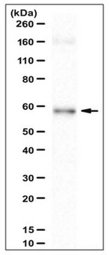
Evaluated by Western Blotting in DLD-1 cell lysate.
Western Blotting Analysis: A 1:5,000 dilution of this antiserum detected CENP-C in 50,000 cell equivalent of DLD-1 human colorectal adenocarcinoma cell lysate.
Western Blotting Analysis: A 1:5,000 dilution of this antiserum detected CENP-C in 50,000 cell equivalent of DLD-1 human colorectal adenocarcinoma cell lysate.
ターゲットの説明
~140 kDa observed. 106.8 kDa (isoform 1) and 61.54 kDa (isoform 2) calculated. The larger-than-calculated target band size is consistent with that reported in the literature (Earnshaw, W.C., et al. (1989). Chromosoma. 98(1):1-12). Uncharacterized bands may be observed in some lysate(s).
物理的形状
Unpurified.
Rabbit polyclonal antibody serum with 0.05% sodium azide.
保管および安定性
Stable for 1 year at -20°C from date of receipt.
Handling Recommendations: Upon receipt and prior to removing the cap, centrifuge the vial and gently mix the solution. Aliquot into microcentrifuge tubes and store at -20°C. Avoid repeated freeze/thaw cycles, which may damage IgG and affect product performance.
Handling Recommendations: Upon receipt and prior to removing the cap, centrifuge the vial and gently mix the solution. Aliquot into microcentrifuge tubes and store at -20°C. Avoid repeated freeze/thaw cycles, which may damage IgG and affect product performance.
その他情報
Concentration: Please refer to lot specific datasheet.
免責事項
Unless otherwise stated in our catalog or other company documentation accompanying the product(s), our products are intended for research use only and are not to be used for any other purpose, which includes but is not limited to, unauthorized commercial uses, in vitro diagnostic uses, ex vivo or in vivo therapeutic uses or any type of consumption or application to humans or animals.
適切な製品が見つかりませんか。
製品選択ツール.をお試しください
保管分類コード
12 - Non Combustible Liquids
WGK
WGK 1
引火点(°F)
Not applicable
引火点(℃)
Not applicable
適用法令
試験研究用途を考慮した関連法令を主に挙げております。化学物質以外については、一部の情報のみ提供しています。 製品を安全かつ合法的に使用することは、使用者の義務です。最新情報により修正される場合があります。WEBの反映には時間を要することがあるため、適宜SDSをご参照ください。
Jan Code
ABE1957:
試験成績書(COA)
製品のロット番号・バッチ番号を入力して、試験成績書(COA) を検索できます。ロット番号・バッチ番号は、製品ラベルに「Lot」または「Batch」に続いて記載されています。
ライフサイエンス、有機合成、材料科学、クロマトグラフィー、分析など、あらゆる分野の研究に経験のあるメンバーがおります。.
製品に関するお問い合わせはこちら(テクニカルサービス)